


West Nile virus infection in a red-tailed hawk
A juvenile female red-tailed hawk was found recumbent and emaciated, with infrequent seizures observed during rehabilitation. The submitting veterinarian collected EDTA whole blood for the West Nile virus (WNV) and Eastern Equine Encephalitis (EEE) virus polymerase chain reaction (PCR) panel and heparinized whole blood for the WNV plaque reduction neutralization test.
As the hawk’s condition continued to deteriorate, euthanasia was elected, and the body was submitted to the Cornell Animal Health Diagnostic Center (AHDC) pathology service for necropsy. Gross examination confirmed severe emaciation with serous atrophy of fat, consistent with the antemortem findings. A brain swab was collected at necropsy for WNV, EEE and avian influenza PCR testing. Molecular testing detected WNV in whole blood and brain swab samples. The PCR Ct (Cycle threshold) value of whole blood was 34.51 and the Ct value of the brain swab was 10.88. As Ct values are inversely proportional to the amount of target nucleic acid present, the lower Ct value in the brain swab indicates a higher viral RNA load in neural tissue, consistent with the known neurotropism of WNV. 1 PRNT yielded a titer of 160, indicating a strong neutralizing antibody response against WNV.
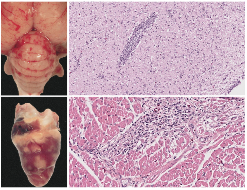
WNV is an arthropod-borne flavivirus capable of infecting a variety of vertebrate hosts, with birds serving as the primary reservoir. The virus is maintained through a mosquito-bird-mosquito transmission cycle, although predatory birds and scavengers may also become infected through ingestion of WNV-infected prey. Clinical signs in avian species may include depression, anorexia, dehydration, and neurologic abnormalities. Post-mortem examination of acute infections may reveal gross lesions of emaciation and hemorrhage, with corresponding histologic findings of cellular necrosis and inflammation in the brain, heart, kidney, spleen, and liver. 2 Sporadic infection of equids and humans occur via mosquito bites and are associated with viral encephalitis. These species are considered dead-end hosts as viremia is insufficient to support transmission back to mosquitoes.
Free-ranging avian species contribute to WNV amplification and the mosquito-driven transmission cycle, highlighting the importance of monitoring avian disease activity to assess public and animal health risks. Active infection can be detected using PCR performed on EDTA whole blood, oropharyngeal or cloacal swabs, or fresh tissue samples including brain, heart, kidney, spleen, liver, and bone marrow.
The AHDC offers multiple antibody tests for WNV, depending on the species of animal. When interpreting the WNV PRNT, paired samples are recommended for an accurate assessment of recent infection versus previous exposure or prior vaccination. However, vaccination is unlikely to be a confounding factor in free-ranging raptors.
References
- Tamba M, Bonilauri P, Galletti G, et al. (2024). West Nile virus surveillance using sentinel birds: results of eleven years of testing in corvids in a region of northern Italy. Frontiers in veterinary science, 11, 1407271. https://doi.org/10.3389/fvets.2024.1407271
- Gamino V, Höfle U. (2013). Pathology and tissue tropism of natural West Nile virus infection in birds: a review. Veterinary research, 44(1), 39. https://doi.org/10.1186/1297-9716-44-39
- Steele KE, Linn MJ, Schoepp RJ, et al. (2000). Pathology of fatal West Nile virus infections in native and exotic birds during the 1999 outbreak in New York City, New York. Veterinary Pathology, 37(3), 208-224. https://doi.org/10.1354/vp.37-3-208