


Two confirmed cases of rabies in a New York beef herd
In September 2026, over the course of three weeks, three adult Angus beef cattle in a herd of 114 animals developed severe acute neurologic signs and died. Clinical signs included excessive salivation and vocalization, absent gaze, running through fences and gates and severe pruritus, leading to self-trauma and skin damage. The owner thought the first animal, a bull, had an esophageal obstruction (commonly referred to as “choke”). The owner passed an orogastric tube to relieve the possible obstruction, but the bull died within hours. The second and third animals (adult cows) became neurologic and died. The owner contacted the herd veterinarian, who collected the appropriate brain sample for rabies testing in both animals. This included a full cross section of the cerebellum and a section of brainstem. The fresh sample must be placed in a sterile container and sent for overnight delivery with ice packs to the New York State Department of Health, Wadsworth Center in Albany, NY.
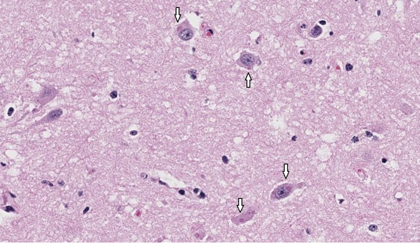
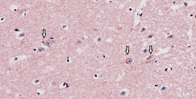
Additionally, formalin fixed tissues and fresh tissues from the third cow were submitted to the Cornell Animal Health Diagnostic Center (AHDC) for histopathology and ancillary testing. A top differential based on the neurologic clinical signs in pasture-based cattle was lead toxicosis. Lead testing on fresh liver tissue was negative. Fluorescent antibody staining for rabies was positive in both cows. Histopathology of the brain performed at the AHDC revealed intraneuronal intracytoplasmic viral inclusion bodies (Negri bodies),1 which is pathognomonic lesion for rabies virus (Image 1). An immunohistochemical stain for rabies was applied to the histology slides as well, staining the Negri bodies a dark red (Image 2).
Rabies virus is a negative sense RNA virus in the lyssavirus genus, Rhabdoviridae family.1 It is zoonotic and causes encephalitis due to neurotropism. It is almost 100% fatal. Rabies is reportable and preventable by vaccination. The remaining cattle on the premise and a single horse were vaccinated for rabies by the herd veterinarian. The exposed farmer was administered post-exposure prophylaxis.
In this case, the veterinarian submitted brain samples directly to the Wadsworth laboratory. The AHDC necropsy laboratory can perform brain extraction on rabies suspect animals from New York and submit the sample to the Wadsworth Center Rabies Laboratory for practitioners as well. Submitting veterinarians must fill out an animal rabies specimen history form in addition to the AHDC submission form. This form accompanies the cerebellum and brainstem samples to the Wadsworth Center Rabies Laboratory.
Reference
- Lahaye X, Vidy A, Pomier C, Obiang L, Harper F, Gaudin Y, Blondel D. Functional characterization of Negri bodies (NBs) in rabies virus-infected cells: Evidence that NBs are sites of viral transcription and replication. J Virol. 2009 Aug;83(16):7948-58. doi: 10.1128/JVI.00554-09. Epub 2009 Jun 3. PMID: 19494013; PMCID: PMC2715764.