

Fibrocartilaginous embolism (FCE) in dogs
Overview
Fibrocartilaginous embolism (FCE) is a condition that affects the spinal cord, most commonly in large-breed dogs. It occurs when a small piece of material from an intervertebral disc enters the bloodstream and blocks the blood vessels supplying the spinal cord, depriving the spinal cord tissue of oxygen and nutrients. The resulting damage leads to a rapid onset of neurological signs, including weakness and paralysis.
Dogs may vocalize in pain at the time of injury, but pain typically subsides quickly and is not a lasting symptom of FCE. There is no specific treatment for FCE. Management focuses on supportive care and rehabilitation. Recovery depends on the severity and location of spinal cord damage. Many dogs show improvement over a few weeks. Some dogs may have a full recovery, but many can have lasting neurologic deficits.
Cause
The exact mechanism of why FCE occurs is not fully understood, but it often occurs during exercise or play behaviors, such as jumping to catch a Frisbee. While FCE is more common in large-breed dogs, it can occur in any breed, typically between three and seven years of age.
FCE occurs when a tiny piece of fibrous cartilage breaks off from the gel-like center of an intervertebral disc and enters the blood vessel that supplies the spinal cord. This fragment blocks blood flow to a portion of the spinal cord, causing a lack of oxygen and nutrients in the area, which leads to tissue death (ischemia) and neurological signs.
Clinical signs
FCE typically causes sudden neurological deficits that vary depending on the location and severity of the spinal cord damage. FCE can deteriorate over the initial couple of hours as more tissue dies, but will become non-progressive and non-painful within minutes to hours.
Clinical signs typically affect one side of the body more severely and may include:
- Loss of coordination or stumbling
- Sudden weakness in one or more legs
- Difficulty walking or complete inability to walk
- Loss of bladder and bowel control as well as deep pain perception in the limbs involved, which is considered the highest degree of severity in spinal cord injury.
Diagnosis
Your veterinarian may suspect FCE has occurred based on a sudden onset of clinical signs and physical exam findings. Advanced imaging, such as MRI, is often used to confirm a diagnosis and rule out other conditions. Referral to a veterinary neurologist may be recommended.
Treatment
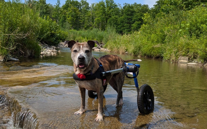
There is no specific medical treatment for FCE. Management focuses on supportive care and rehabilitation. Your veterinarian may refer you to a rehabilitation specialist.
Supportive care may include:
- Providing soft bedding and frequent repositioning to prevent pressure sores
- Assisting with mobility using slings or harnesses (e.g., the Help ‘Em Up® Harness)
- Manual bladder expression if the dog cannot urinate independently
- Monitoring for urinary accidents and keeping the dog clean and dry to prevent skin irritation
Rehabilitation may involve:
- Passive range of motion exercises
- Assisted standing and walking
- Massage and stretching exercises
- Physical therapy exercises
- Underwater treadmill therapy
- Neuromuscular stimulation
Outcome
The prognosis of FCE depends on the severity of spinal cord damage. Many dogs begin to show improvement within five to fourteen days. Some dogs may recover fully, though it is often more common to have some lasting neurological deficits. With appropriate care and rehabilitation, many dogs with FCE can return to a good quality of life, even if some neurologic deficits persist.
A poorer prognosis is associated with:
- No deep pain perception at the time of diagnosis
- Symmetrical clinical signs (which indicate both sides of the cord have been affected)
- Signs of lower motor neurons involvement (loss of reflexes and tone in the limbs and or tail)
- No improvement within the first two weeks
This page was last updated on Tuesday, Dec 09, 2025