


Project Overview
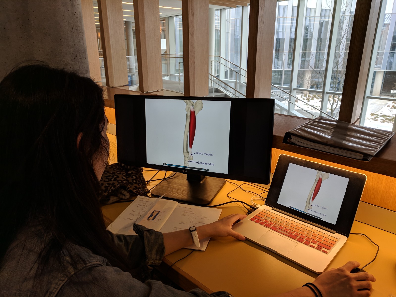
The Equine Carpus Modules provide students with interactive learning materials that provide the basics of the bony anatomy, soft tissues, and radiography of the carpus. This knowledge develops as students watch videos, view pictures and radiographs, and perform virtual diagnostic tests within the framework of two real-world cases.
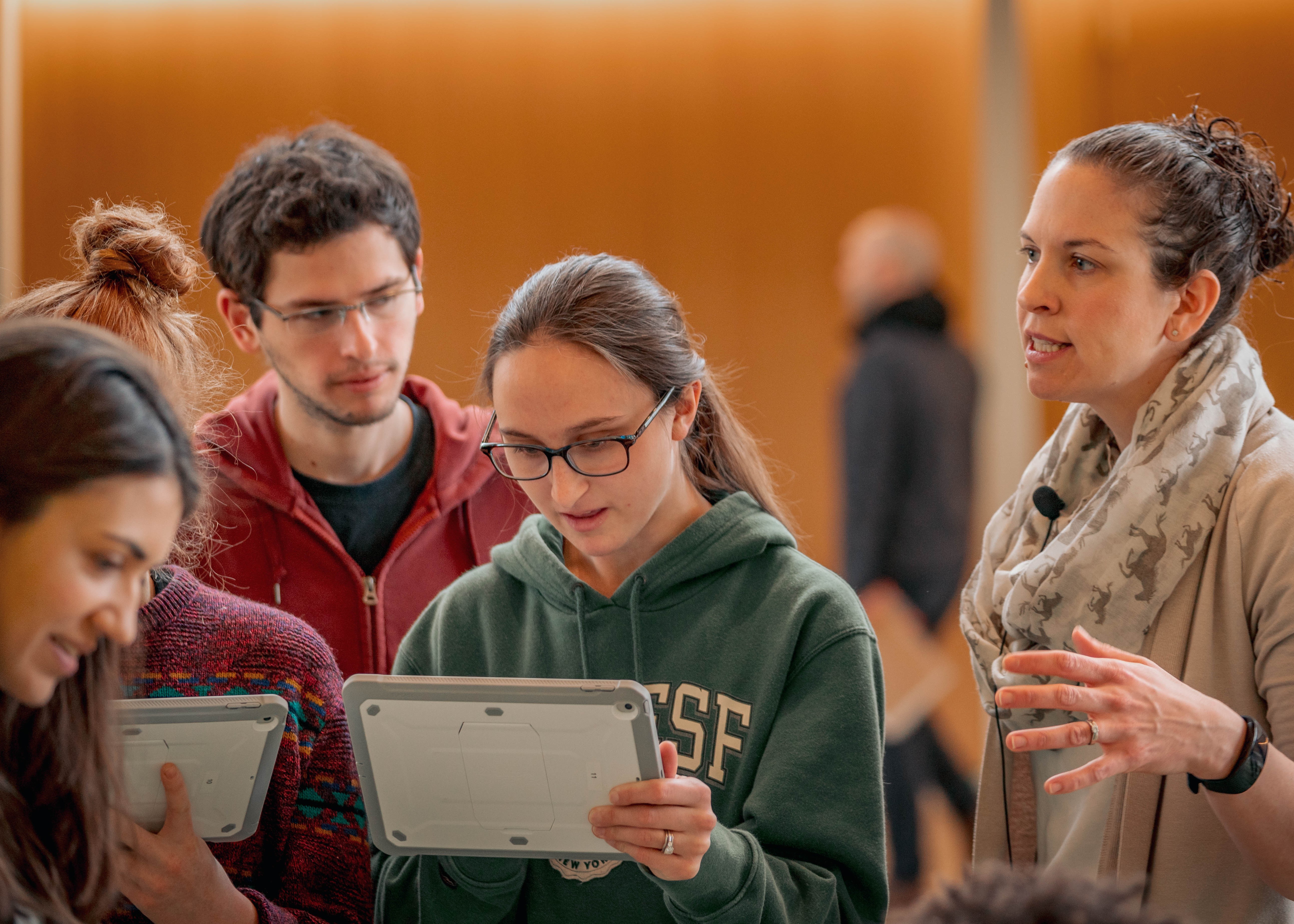
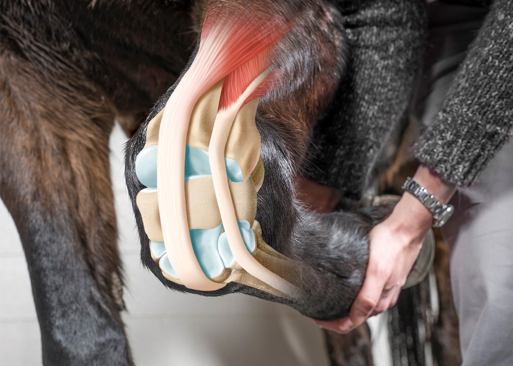
Developed by Dr. Allison Miller and the Educational Support Services team (ESS), the carpus modules ask students to explore a 3D carpus representation; while rotating, flexing and viewing different layers of tissue, students build a conceptual map of the region. Students apply this 3D understanding of the anatomy to real-world cases that are completed as an in-class group activity. This activity allows students to develop clinical reasoning and decision-making skills as they directly apply pre-clinical coursework in a virtual clinical context. This experience culminates with the use of an Augmented Reality (AR) application on an iPad and allows students to practice a notoriously difficult concept to learn: visualizing and virtually capturing five different radiographic views of the equine carpus.
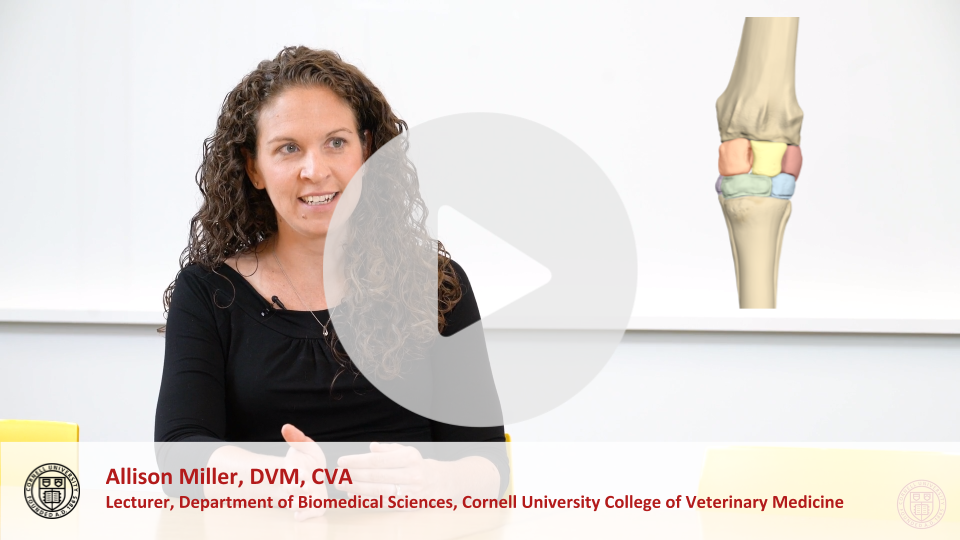
“When I first started this project, I imagined it simply as an additional online resource that students could use outside of class time on their own time without much direction. What evolved from multiple brainstorming meetings was beyond anything I could have imagined. The technology appeared to increase student understanding of the equine thoracic limb. Students came to dissection lab with more knowledge and appeared to me to more efficiently approach, and finish their dissection. The knowledge seemed to stick as they applied it to the in-class cases. Walking around the classroom during the in-class activity, I heard such brilliant conversations as they drew on their knowledge from the out-of-class activity and their dissection labs. Best of all, they seemed excited and engaged with the material when it was presented in this way as compared to a didactic lecture.”
-Allison Miller, DVM, CVA
Student Reflections
- I really liked this module-- it made limb anatomy seem much more approachable and the information that was included was relevant and concise.
- I think it’s a really cool program and helpful activity!
- Overall, I found these modules very helpful, and I thought they were a great way to learn the material.
Project Support
This project was made possible through an Educational Technology Innovation Grant from the Cornell University College of Veterinary Medicine.