Ventilator breathes new life into patient
The patient arrived at the hospital in very bad shape. Her breathing was labored; her body temperature was well below normal. “She was near death on presentation,” said an observer. “She had deteriorated to the point where either she was going to die or we would have to take over and breathe for her.”
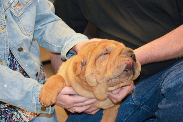
So Shiloh, a six-month-old Shar Pei puppy, was put on a ventilator and admitted to the Intensive Care Unit at the Cornell University Hospital for Animals (CUHA). She was treated for severe pneumonia. A week later, Shiloh bounded into the arms of her owners and went home, wagging her tail behind her.
Few veterinary hospitals have ventilators. They are expensive, and attending staff need special training to monitor the patient and perform round-the-clock adjustments as the patient’s condition changes. At Cornell, residents and faculty train on a simulator that mimics real spontaneously breathing patients with unhealthy lungs and complications of ventilation, a gift from the Triad Foundation and the Hatfield Family. “The simulator offers insights into novel ways of managing patients, gives you confidence that you can manage complications that you would only see rarely in patients,” says Dr. Robert Goggs, who headed Shiloh’s medical team.
Shiloh, who was referred to Cornell by her regular vet, did not respond well to initial attempts to stabilize her breathing and was put on the ventilator as an emergency measure. A CT scan revealed extensive damage from accumulations of fluid in the lungs. Shiloh’s lungs were so severely affected that they could no longer support her and needed time to heal. The ventilator became the primary means of treatment, taking over the work of breathing so the puppy’s lungs could repair. A subsequent test revealed pneumonia, and antibiotics were added to her treatment plan.
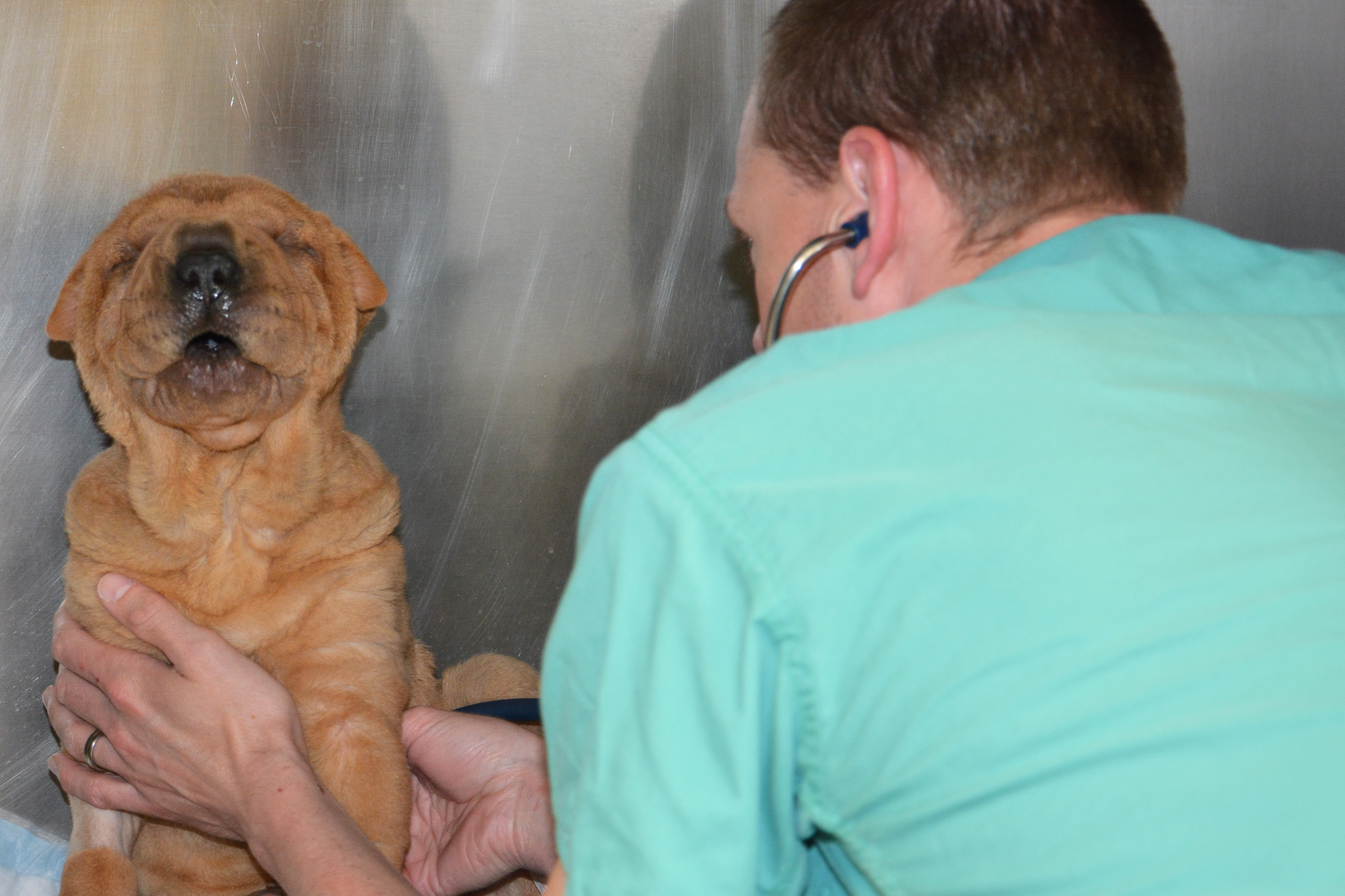
The ICU staff monitored Shiloh 24 hours a day, adjusting the settings to make sure her blood was adequately oxygenated and watching for the complications associated with the ventilator itself.
“Mechanical ventilation is an unnatural way of breathing,” explains Dr. Goggs. “Normally we suck air in, but the ventilator pushes air in. Damaged lungs can overdistend as air is pushed in under pressure.” On the second day, Shiloh developed a pneumothorax - air leakage into the pleural space (between the lungs and the chest wall), a serious complication of mechanical ventilation.
The team adjusted the ventilator settings with each change in Shiloh’s condition to avoid an additional complication: The muscles that regulate breathing can weaken when they aren’t used, making a patient more dependent on continued ventilation. Gradually reducing ventilator support encourages those muscles to take on more of the work themselves.
By the morning of the fourth day Shiloh needed so little support from the ventilator that she was weaned from it. By that evening, she was eating well and wagged her tail when her owners came to visit. Six days after arriving at CUHA she went outside to play in the sunshine, and on the eighth day she went home. Shiloh returned for a follow-up appointment later that week and showed continued improvement.
Dr. Goggs doesn’t think Shiloh will experience severe lasting effects from her illness. “A little emphysema, a little scarring perhaps, but it’s unlikely and won’t be clinically significant.” He emphasizes that Shiloh was a lucky puppy: “It’s fortunate that she was a healthy young dog before this crisis. And we are fortunate to have a very capable ventilator and a fantastic team of people who always go the extra mile for their patients.”
Published May 13, 2015