Cornell researchers uncover hidden player in gut growth
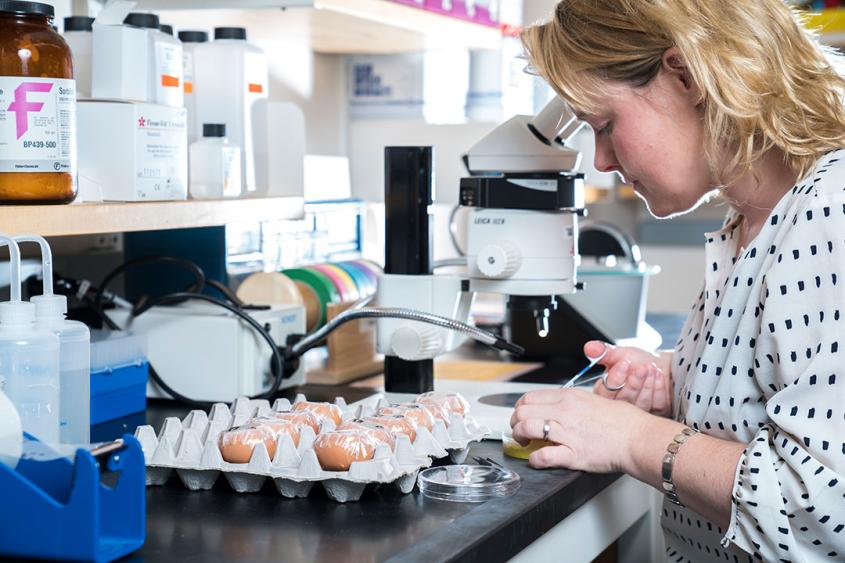
Dr. Natasza Kurpios, associate professor in the Department of Molecular Medicine, working in her lab. Photo by the College of Veterinary Medicine.
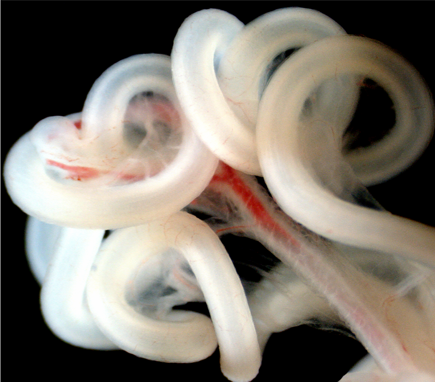
A previously ignored part of the intestine has turned into the key to its most crucial moment in embryonic development: the rotation that winds the small and large intestine into its familiar twisted form.
Where this rotation is triggered and by what is the subject of new research from Dr. Natasza Kurpios’ lab in the College of Veterinary Medicine, which shows that the right side of the intestine’s connective tissue triggers the gut’s change from a symmetrical tube to coiled spaghetti. This is a major shift in the field, which widely believed the trigger occurred on the left.
“On the outside, human bodies appear symmetrical, but on the inside, organs like the heart, liver, lungs and stomach are all asymmetrical,” said Kurpios, associate professor in the Department of Molecular Medicine and senior author on the paper, which published today in Developmental Cell. Asymmetrical organs are found on just one side of the body, or across both left and right but with their components distributed unevenly. The Kurpios lab studies this left-right asymmetry in organ development.
Pinpointing when and how gut rotation begins lends insight into embryonic development and lays the groundwork for clinical therapies. “If we can identify some of the critical pathways that regulate left-right asymmetry in the digestive tract, then we can expand those studies to more complex organs like the heart,” said Aravind Sivakumar, Ph.D. ’17, first author on the paper and former graduate student in the Kurpios lab.
The embryonic intestine itself is comprised of a gut tube and connective tissue. The gut develops into the small and large intestine, and the connective tissue, or dorsal mesentery, not only suspends the gut from the dorsal body wall but also provides all vasculature like arteries and lymphatics to the intestines. Previous research, some of which was conducted at Cornell, demonstrated that it was the mesentery that caused the rotation of the gut and not the other way around. This rotation is predictable in healthy embryos – always counter-clockwise and always in the same pattern.
“Left-right asymmetries in the intestines are quite concealed in humans because the gastrointestinal tract is huge, but its development is very carefully choreographed,” said Kurpios.
While connected, the left and right sides of the mesentery are discrete. For many years, biologists have focused on the gene Pitx2 found within the left side, thinking that it was the key to understanding abnormal gut rotation cases, such as congenital birth defects. The potential of Pitx2 and its assumed role in triggering gut rotation accelerated research into the left side of the mesentery, leaving the right side mostly unexplored.
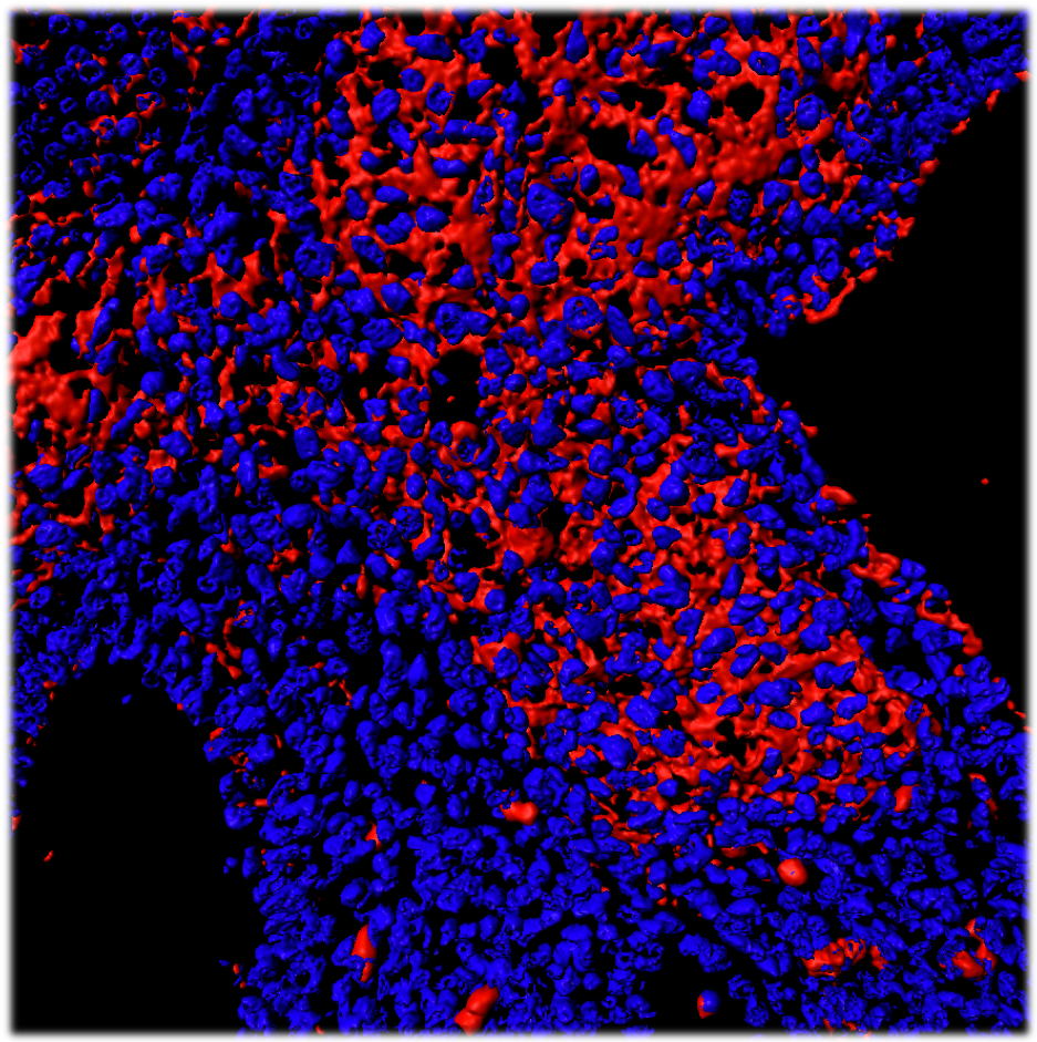
The Kurpios lab’s new research pinpoints that this trigger not only happens on the right side of the mesentery, but also several developmental stages earlier than noted on the left.
“We found that it’s the right that expands and collectively swings the gut tube on its side many embryonic stages before we see statistically significant movement on the left,” said Sivakumar.
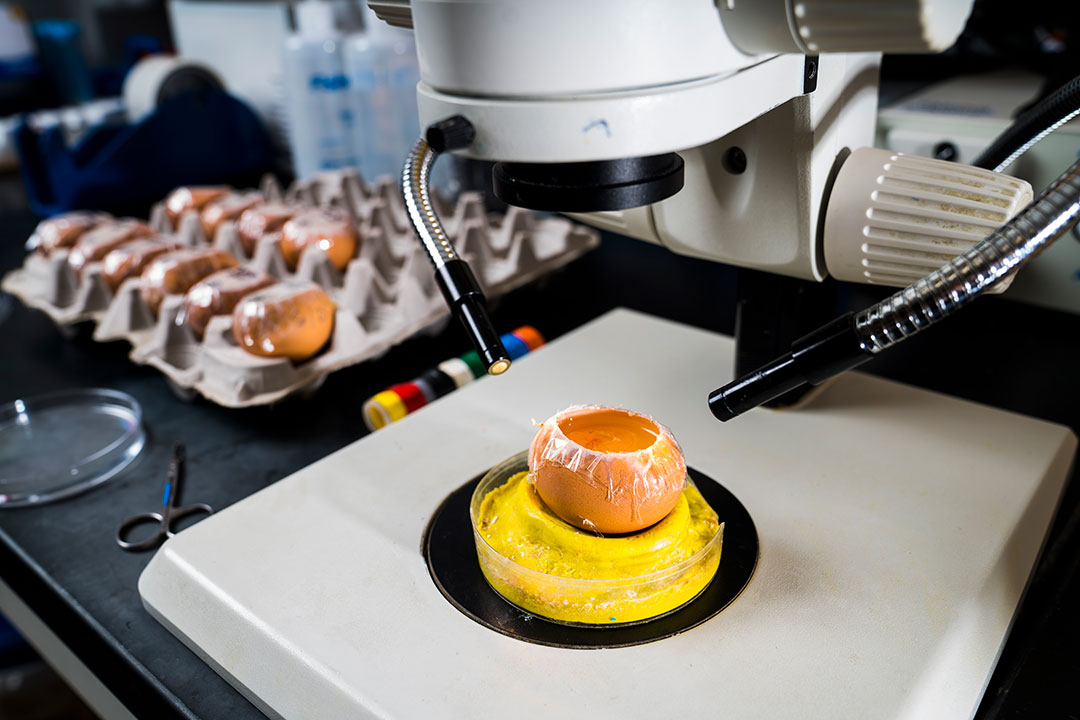
Using chicken and mice models, Sivakumar traced the cause to a surprising source: a simple polysaccharide known as hyaluronan (HA), also widely used in wrinkle cream for its hydrating properties. Upstream from the gut, the enzyme Tsg6 covalently modifies HA, which spurs the expansion of the right side and thus the rotation.
“The opinion in the field was that the right side is passive, that it was just along for the ride,” said Kurpios. “What Aravind found completely changes the way we view left-right symmetry and puts the right side on the map.”
This work was supported by the March of Dimes; National Institute of Diabetes and Digestive and Kidney Diseases; the National Institute of Environmental Health Sciences; and the National Heart, Lung, and Blood Institute Program of Excellence in Glycosciences. Aparna Mahadevan, Ph.D. ’16, Mark Lauer, Ricky Narvaez, Siddesh Ramesh, Cora Demler, Nathan Souchet, Vincent Hascall, Ron Midura Stavros Garantziotis, David Frank and Koji Kimata also contributed to the study.
By Melanie Greaver Cordova