Laying the groundwork to find new therapies for oral tumors
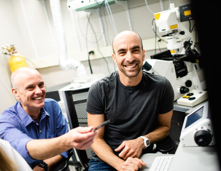
Dogs with a type of mouth cancer called oral squamous cell carcinoma (OSCC) often undergo a disfiguring surgery that sometimes removes up to a quarter of the jawbone. Drs. Santiago Peralta and William Katt think there might be a better way. They are developing new models of OSCC with funding from the Cornell Richard P. Riney Canine Health Center to test out novel therapies in the lab that may one day be used in patients.
"We deal with this type of tumor routinely in the clinic," said Peralta, an associate professor of dentistry and oral surgery at the Cornell University College of Veterinary Medicine. "Treatment options have been unchanged for a very long time and the surgical solutions are often devastating to the patients. With the advent of novel technologies and amazing expertise from Cornell, we realized we could join forces and come up with better ways to treat OSCC patients."
Katt, a senior research associate in Molecular Medicine at Cornell specializing in cancer drug discovery, met Peralta at a cross-campus conference. They decided to team up and apply their very different areas of expertise to treating oral tumors.
Even for humans with mouth cancer, there aren't many "gentle therapies," said Katt. "Canine oral cancer is thought to really well represent human oral cancer. So, we hope that anything we discover that works in canines could then be adapted to help human patients as well."
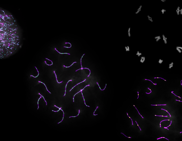
Scientists must screen potential new drugs in the lab for safety and effectiveness before giving them to patients. But for canine oral tumors, there are no screening tools available. To correct this problem, Peralta is taking cancerous tissues from his patients and growing them inside mice, while Katt is turning them into cell lines on plates in the lab. They aim to develop up to 10 mouse models with matching cell lines to represent the genetic diversity found in OSCC tumors.
Next, the team will test potential drugs in the cell lines and then in mice implanted with the tumors to identify ones that successfully shrink them. Ultimately, they hope to discover effective pharmacological approaches that can be tested further in clinical trials to treat dogs with OSCC. The work may one day result in fewer disfiguring surgeries.
Additional collaborators on this project include Rishi Puri, project manager of the PATh PDX Facility, which is developing the mouse tumor models; Jen Grenier, director of the Genomics Innovation Hub, who is helping with tumor sequencing efforts; and Gerald Duhamel, professor of anatomic pathology in Biomedical Sciences, who confirms that the tumors are in fact OSCC. Susan Garrison and Denise F. LaLonde-Paul from the Cornell Veterinary Biobank are helping to facilitate the collection and storage of samples.
"We are making excellent use of the Cornell facilities and the experts they have on hand to make sure everything is being done correctly," said Peralta. "This is critical for discovery."
Written by Patricia Waldron
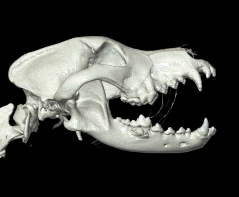
The dog in this image from a CT scan has an oral squamous cell carcinoma (OSCC) on its lower jaw (left). Surgery to eliminate the tumor required the removal of a substantial portion of the jaw (right).
Credit: Images courtesy of Santiago Peralta