Assessment of vision and common eye problems in horses
This is an accompanying article to a Cornell Equine Seminar presented on Mar. 15, 2022 by Dr. Kelly Knickelbein, assistant clinical professor in the section of ophthalmology.
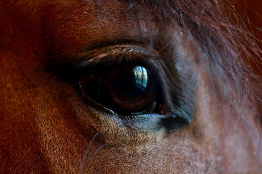
Horses are thought to have good vision – but their large eyes are vulnerable to injury and diseases that can result in the loss of eyesight if they are not properly cared for. Board-certified ophthalmologist Dr. Kelly Knickelbein explains the basics of equine vision, common diseases, and how best to care for your horse’s eyes.
What follows here is an overview. Please watch Knickelbein’s entire presentation for more extensive information.
How the equine eye works
“Vision is an incredibly complex, physical and biochemical process, through which light that enters the eye is made into an image by the brain,” Knickelbein says. The incoming light reflected from objects in the environment is bent by the cornea and the lens and hits the retina, the sensory tissue in the back of the eye. The optic nerve, the optic chiasm (the part of the brain where the optic nerves cross), and the optic tract (a bundle of nerve fibers coming from the optic chiasm) carry these signals to the visual cortex of the brain, where they are integrated into an image that can be perceived by the animal.
This multipart process is further complicated by the fact that the eyes are often in motion – controlled by the extraocular muscles – and that some of the information taken in by the eye is transmitted to the opposite side of the brain.
Light perception
Horses are generally thought to have good vision. Their large eyes and big pupils take in a lot of light, and the size of their retina allows a high number of cells to be involved in light capture and processing. Among these photoreceptors, rods – responsible for vision in dim light – greatly outnumber cones, which are responsible for bright light vision as well as color perception. (The ratio lies somewhere between 9:1 and 20:1, according to the literature.)
The tapetum lucidum – the reflective structure at the back of the eye in horses and many other species, but not humans – further increases the light-capturing ability of the photoreceptors by bouncing light back towards them a second time, improving night vision.
Because horses spend most of their time outdoors and are also exposed to bright conditions such as midday sun, two mechanisms protect the retina from photic damage and prevent overstimulation of the cells to allow vision to continue: The pupil can constrict to significantly limit the amount of light entering the eye, and the corpora nigra – bulbous structures protruding into the space of the pupil – acts as a kind of shade.
Field of vision and motion detection
“One thing that's particularly impressive about horses’ vision is that they essentially have a panoramic field of view,” says Knickelbein. While their binocular vision, or what they can see simultaneously with both eyes, is somewhat limited at about 65 degrees, they can see far in front and behind with each individual eye. Their focus is crispest in the center and fades out peripherally. “This makes sense, because horses are prey animals, so they need to be able to detect predators anywhere in their surroundings, including off in the distance behind them, and this acute view of the horizon line lets them detect small movements in the distance.” Blind spots, on the other hand, exist at short distances directly in front of and behind the horses.
Depth perception
The integration of different images perceived by the left and right eyes is a critical component of depth perception. Nevertheless, horses with unilateral blindness (loss of vision in one eye) do compete successfully in equestrian sports. One example is Viscera, a Hanoverian mare who competed for Team Sweden in Eventing at the Tokyo Olympics. “The fact that she maintains her ability to compete at the highest level of the sport, despite being unilaterally enucleated [blind], indicates that she has cues from her remaining eye allowing her to perceive depth,” Knickelbein says.
Color perception
Horses have two types of cone photoreceptors in the retina, compared to three in most humans (some people have only two). They are sensitive to the blue wavelength and to one that lies between green and red. As a result, horses see color, but on a smaller spectrum than humans typically do.
Assessment of vision in horses
A vision assessment begins with gathering a thorough history from those who regularly care for the horse.
Questions cover:
- use of the horse: e.g. eventing or dressage
- environment: e.g. outdoors or stalled
- diet
- concurrent health conditions and medications that may impact treatment recommendations
The veterinarian will then observe visual behavior from afar to pick up clues about how the horse is seeing and perceiving its surroundings, both in well-lit and dimly lit conditions.
A neuro-ophthalmic examination tests responses to visual stimuli, reflexes to light and touch, and pupil size and movement. Most very young foals will not yet close their eyes in response to a hand moving towards them, but they learn quickly to do so for protection.
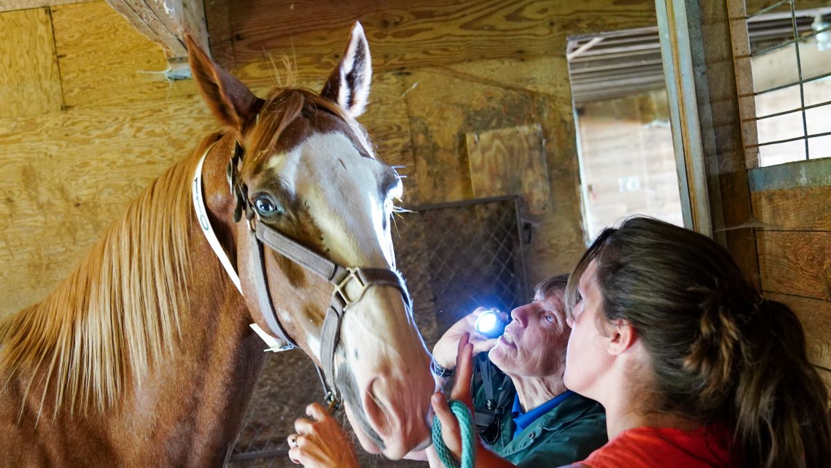
The ophthalmic examination with a variety of lighting and magnifying instruments determines whether any structural abnormality of the eye could be compromising vision.
Advanced diagnostics
Occasionally, horses have a normal eye exam, but their history or behaviors suggest that they are not seeing well. In this case, veterinary ophthalmologists reach for advanced diagnostics such as refraction, which uses a light source and a series of lenses to determine if there is an optical refractive error in the eye – the equivalent of what the horse’s glasses or contact lens prescription would be, and a relatively rare condition.
An electroretinogram, or ERG, is another advanced test. It checks the electrical function of the retina to verify whether the horse’s photoreceptors and adjacent cells are functioning properly. In the case of congenital stationary night blindness (inherited in the Appaloosa and a few other breeds), for example, it may show that the photoreceptors are working but not transmitting the signal to the adjacent cells in the retina to allow for the photo transduction process to continue and vision to occur.
Common eye problems that impact vision
Among the most common eye problems in horses are:
- corneal disease (ulcers, abscesses, immune-mediated keratitis)
- uveitis
- cataracts
- squamous cell carcinoma
- corpora nigra cysts
They will be discussed in more detail below.
Corneal ulcers
Horse eyes are quite prone to trauma because they are large and situated on the sides of the animal’s head. Even simple, superficial scratches can quickly become infected with bacteria or fungal organisms and lead to severe inflammation. To preserve the integrity of the eye, it is therefore important to identify ulcers – the loss of the top layer of corneal cells – early. Ulcers can be diagnosed with fluorescein stain, which sticks to the deeper layers of the cornea, indicating that the top layer of cells is no longer present.
Horses with superficial ulcers often experience good outcomes with only mild scarring unlikely to make a significant impact on their vision. Animals with severe corneal infections often have much more severe scarring, whether or not they are able to be managed medically or surgically. Some horses may lose vision due to a severe corneal infection, and this can lead to the need to surgically remove the eye.
Corneal stromal abscess
Corneal stromal abscesses typically develop due to microtraumas to the cornea that inoculate infectious organisms (fungus or bacteria). Because the top layer of cells is intact, fluorescein stain is negative with this condition.
Such cases can experience significant ocular pain and may require surgery to remove the infection. The infection may also move inside of the eye and cause severe inflammation. In the worst case, a horse may need to have its eye removed if vision is lost and pain cannot be controlled.
Immune-mediated keratitis
Immune-mediated keratitis describes a non-infectious inflammation of the cornea. The condition can lead to the cornea becoming opaque (due to infiltration of white blood cells and blood vessels), and it may or may not cause discomfort in affected horses.
Topical medications are the first line of treatment, while implants can also help to control the disease. Some horses will ultimately need to undergo surgery to remove the affected part of their cornea to limit the opacity and its impact on vision. The surgery itself will result in some scarring.
Uveitis
Uveitis is inflammation of the uveal tract, the vascular layer of the eye. “It’s the damage to the other structures in the eye from that inflammation that leads to visual impairment,” Knickelbein explains. For example, a horse may develop adhesions of its iris to its lens, limiting the ability of the pupil to move. Cataracts (opacification of the normally transparent lens), is another vision-impairing long-term effect of uveitis.
Equine Recurrent Uveitis, a complex disease commonly referred to as “moon blindness,” is the most common cause of blindness in horses. It is thought to be an immune-mediated disease with known genetic and environmental/infectious risk factors.
“One of the most devastating things about the disease is that some horses can have chronic inflammation for years without any outward signs,” says Knickelbein. “Oftentimes we see horses for the first time when visual impairment is noticed, because they've actually gone completely blind in one or even both of their eyes.”
Corpora nigra cysts
Corpora nigra cysts are fluid-filled circular structures extending from the corpora nigra of the iris. While they do not cause harm to the eye, they can obscure vision.
“This is actually quite a satisfying disorder to diagnose, because we do have an effective treatment for it,” Knickelbein says. A laser can be used to create small holes in the cyst, deflating them.
Cataracts
Cataracts are a frequent cause of visual impairment in horses. They can occur on their own or, most commonly, as a consequence of chronic uveitis. Cataracts can be removed surgically, but eyes with uveitis are not good candidates.
Squamous Cell Carcinoma
Squamous cell carcinoma is the most common cancer to affect the eyelid and ocular surface in horses. Several breeds, including Haflingers and Belgians, are genetically predisposed to developing this cancer.
Treatment depends on the size and location of the tumor. Options include surgery, chemotherapy, cryotherapy, and radiation therapy, amongst others. In some cases, removal of the eye is advisable to try to prevent spread to neighboring tissues.
This tumor type is highly invasive and tends to recur. “So really close monitoring is indicated, no matter where it arises from or how long ago it occurred,” says Knickelbein. “In any horse that’s been diagnosed with and treated for squamous cell carcinoma, we recommend having eye exams at least twice yearly, as well as the use of a good UV-blocking fly mask.”
Tips for keeping your horse’s eyes healthy
Knickelbein suggests looking at your horse’s eyes daily, monitoring for:
- discharge
- squinting, eyelid closure
- cloudiness, opacity
- eyelid swelling
“It's important that you act on any changes to the eyes really quickly by calling your primary vet or
our service to schedule an evaluation, because a lot of ocular signs are nonspecific,” Knickelbein says. “Excessive tearing from the eye could be anything from a simple blocked nasolacrimal duct – not an emergency but something that should be dealt with – to chronic uveitis that is just now manifesting and may still be treatable. Acting quickly can potentially save their vision.”
She also recommends fly masks that protect horses from flies, wind, allergens, and the sun. UV rays are a major contributor to the development of ocular squamous cell carcinoma. Breeds with a genetic predisposition to squamous cell carcinoma (Haflingers, Belgians, Rocky Mountain Horses, among others) as well as horses with non-pigmented skin around their eyes should wear fly masks with at least 90 percent UV protection. Take off the mask at least once daily to check the eyes for any concerning changes.
Managing blind horses
Horses adjust to loss of vision on an individual basis. It is important to keep blind horses safe in their environment – whether a stall, paddock, or pasture – without obstacles that may harm them.
 Help your horse with a consistent routine with the same handlers, feeding times, placement of water buckets and feed, and companions. “They are herd animals, so blind horses can do well with a kind of buddy horse to assist them in using their other senses to navigate the world,” Knickelbein explains. Tips include selecting a calm buddy horse that does not startle easily, putting a bell on the buddy if vision loss is a recent change, and using verbal cues and touch to interact with the blind horse.
A visit to the ophthalmology service
If your horse will be best served by coming to the hospital for an evaluation, you will usually be referred to the ophthalmology service by your primary veterinarian. Each case receives a complete ophthalmic exam and individualized recommendations, based on the diagnosis and the needs of the horse and owner.
Some of the advanced diagnostics available at the Cornell University Hospital for Animals (CUHA) include high-frequency ultrasound for a detailed view of the intraocular structures; in vivo confocal microscopy to investigate corneal infections and diagnose immune-mediated keratitis; as well as electroretinography.
CUHA’s ophthalmologists are trained in advanced surgical techniques, including corneal and intraocular surgeries such as cataract surgery. “One of the really nice things about being at Cornell is the ability to work closely with other specialists, including our equine medicine team, the equine surgery team, and our anesthesia service,” Knickelbein adds.
Treatment decisions happen in partnership with owners, and referring veterinarians are updated routinely to ensure the best continuing care. “We hope to provide comprehensive care alongside your primary veterinarian,” Knickelbein says.
Watch Knickelbein’s full talk online, and see the entire Cornell Equine Seminar Series videos here.