Equine pre-purchase exams: What to expect

This is an accompanying article to a Cornell Equine Seminar presented on Apr. 18, 2023 by John Pigott, D.V.M. ’09, associate clinical professor and clinical director of Cornell Ruffian Equine Specialists.
Pre-purchase exams by qualified veterinarians take some of the guesswork out of buying a new horse. Dr. John Pigott, a specialist equine veterinary surgeon, walks us through the process and explains what to do with the information you learn. What follows here is an overview. Please watch Pigott’s entire presentation for more extensive information.
Goal of the pre-purchase exam
A pre-purchase exam is a single-day assessment of the overall health and soundness of a horse at the time of the exam. Rather than giving a passing or failing grade, veterinarians evaluate the risk associated with the purchase for the animal’s intended use. They will not be able to make any guarantees for its future health. There are many reasons — such as past personal experiences or a horse’s proven performance record — why buyers may decide to make a purchase even with risky conditions or pass on an animal despite a clean bill of health.
Pre-visit conversation
Before the pre-purchase exam, a conversation about expectations, costs, use of the animal and relevant tests is very important. While the veterinarian ultimately works for the buyer, a trainer or agent may act as their representative, so everyone should be made aware of how communication will work. Sometimes a non-local buyer may also want to consult a trusted veterinarian closer to home, who will receive relevant videos and radiographs for evaluation.
Identifying mismatches
Pigott generally considers it a trainer’s or agent’s job to point out mismatches between a horse and a potential buyer. “But sometimes there's a clear mismatch and I will make people aware of my opinion, but it has to be a pretty significant case,” he says.
History
The first step in a pre-purchase exam is to gather as much information as the seller will provide about the horse. (The legal requirements for disclosure at the time of pre-purchase vary by state.) Pigott will note any medications, the level of work over the past six months, any lameness issues, and dates of recent routine healthcare. If he has previously treated the horse under consideration — this may happen when horses are sold within barns under the same trainer, for example — Pigott asks the seller for permission to divulge the medical history. “If a seller does not disclose, it creates a real sticky situation,” Pigott says. To avoid any ethical dilemmas, it can help to find a neutral veterinarian without any previous connections to the animal.
Examination
For a reliable examination, find a practitioner who is detail-oriented and has some experience. (Not all veterinarians are willing to do pre-purchase exams because there can be “a lot of headaches” involved, Pigott says.)
 During the exam, the veterinarian checks all the major body systems for abnormalities and risk factors for potential future issues, including:
- cardiothoracic: murmurs, arrhythmias, nasal discharge
- dermatologic: areas of depigmentation, masses, dermatitis, surgical scars
- oral: dental abnormalities
- ophthalmic: cataracts, uveitis, corpora nigra cysts
- musculoskeletal: lameness, conformation, gait
- neurologic: normal reflexes, muscle atrophy, coordination
- reproductive: breeding soundness (done by theriogenologists), masses, discolorations
Based on the findings, the veterinarian may suggest additional tests or a second opinion from a specialist.
More details on each part of the exam follow below.
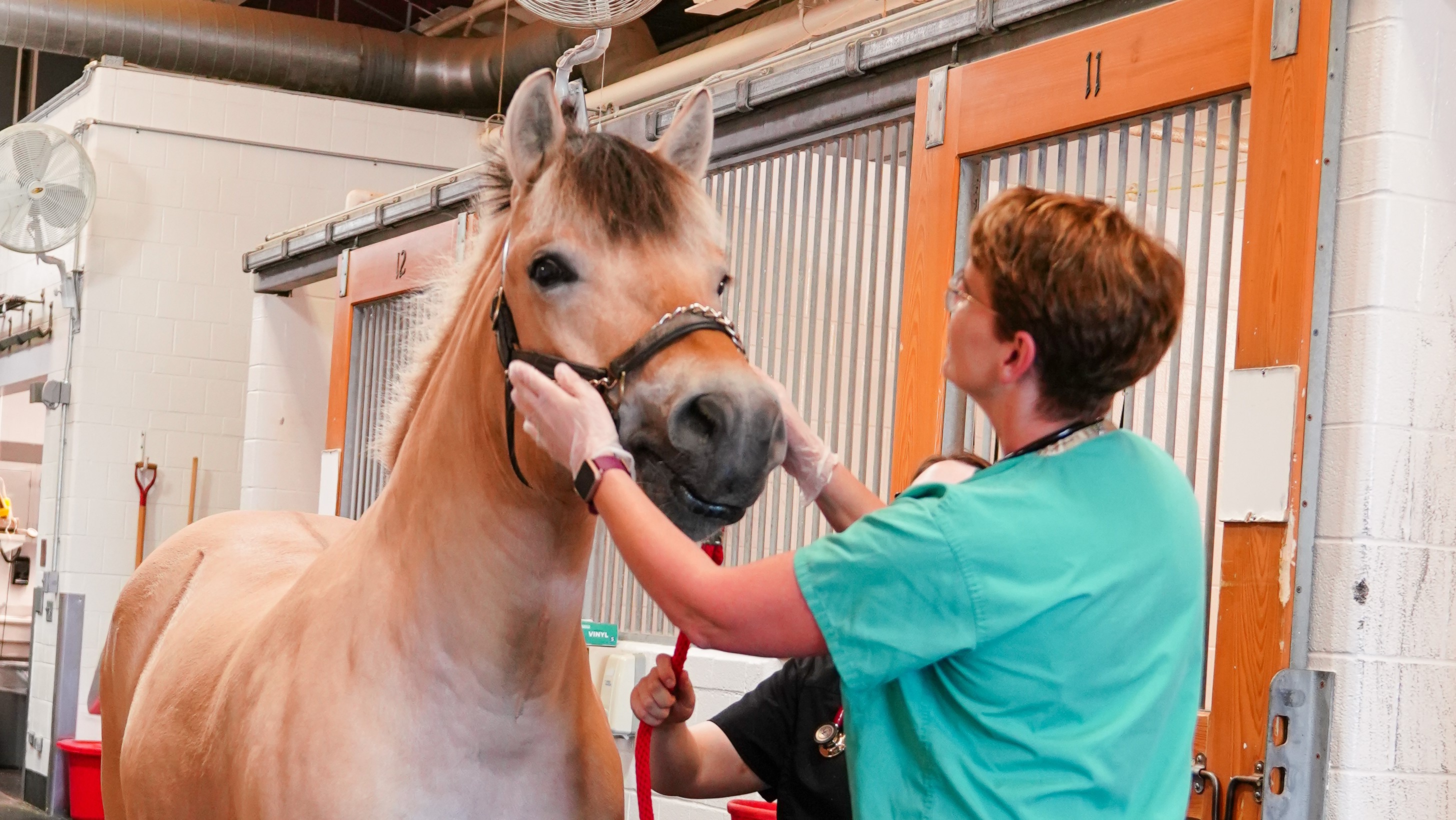
Physical exam
First, Pigott takes a stethoscope to the horse. If he hears anything abnormal in the heart, he may suggest an ultrasound of the heart or an electrocardiogram to the buyer.
Next, he looks for muscle atrophy, which can be a sign of previous injury, or – if it is one-sided – of past equine protozoal myeloencephalitis (EPM). He also palpates for muscle asymmetry in the larynx and listens for noises in exercise that can give clues to underlying issues.
Dermatologic exam
Surgical scars can be easily overlooked in some horses, especially if they are not clipped. Pigott looks for ventral abdominal scars from colic surgery, which may be hard to spot, depending on how the body wall healed. He will also search for any scarring from a palmar digital neurectomy – a procedure that removes a section of nerve from the foot – or a fasciotomy and neurectomy for high suspensory injuries. A tie-back scar at the throat would be a reason to scope the airway to check for previous throat surgery for a roarer.
The significance of masses depends on their location and how they look. “The big masses we’re looking out for are sarcoids,” Pigott says. This type of skin tumor can remain dormant for many years and suddenly activate to grow quickly. Other tumor types include squamous cell carcinoma and melanoma. Masses in and around the third eyelid, on the cornea of the eye, around the shaft of the penis, or over joints or a tendon sheath are all red flags for Pigott. Any tail rubbing could be evidence of allergies but also of parasites.
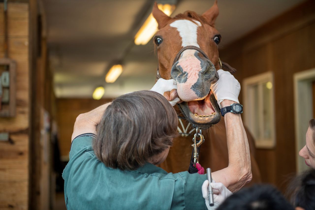
Oral exam
Pigott generally performs unsedated oral examinations, looking at wear patterns and for missing or broken teeth. Some abnormalities in the back of the mouth can be difficult to see. If he has any concerns, he requests a sedated exam with the owner’s permission.
Ophthalmic exam
Eye exams generally take place without dilation, but the veterinarian then has limited visibility of the back of the eye.
Pigott looks for signs of uveitis (inflammation inside the eye) as well as corpora nigra cysts, which may obstruct vision, and cataracts.
Musculoskeletal exam
During the musculoskeletal exam, Pigott checks conformation, the overall structure – for example whether the horse toes in or out or whether it is upright through the hind end. Some types of conformation are more prone to specific injuries. The animal’s back, overall muscling, and top line also convey important information about its suitability for certain activities.
Club feet, if not severe, can often be managed with appropriate trimming and shoeing. Horses with this condition frequently land toe to heel, causing increased stress in the coffin bone.
Heel shearing can have several different causes. It may be evidence of lameness, the horse’s chronic asymmetric growth, or inappropriate trimming by a farrier. “So we have to figure out what is manageable and what isn’t,” says Pigott.
He looks closely at the type of shoes on the horse’s feet and their wear patterns. A shoe that is wider on one side supports that part of the foot – perhaps due to an injury. Glue-ons are popular among hunters for being lightweight, but they are also used on horses with chronically poor hoof walls that can’t take a nail. “Those would be more expensive to maintain in the long run,” Pigott explains.
Using a hoof tester he identifies bruising on the foot and looks for quarter cracks. He can later relate any patterns of sensitivity to the horse’s behavior during the dynamic exam that follows.
By palpating every aspect of the horse’s body, particularly over the joints and tendon sheaths, Pigott finds spots of extra fluid. Such effusion may be a sign of pathology but could also occur in an older horse working hard. “Windpuffs” (fluid in the tendon sheath) on both legs are less concerning than fluid on only one side.
Pigott always palpates areas of thickening. Thickened tendons or ligaments may represent previous surgery – for example, check ligament surgery as a younger horse for club feet – or prior injury. He always offers ultrasound of these tissues.
Smooth, non-painful splints (bony enlargements) don’t bother Pigott, who worries more about larger bony calluses. These could be a blind splint that goes towards the suspensory ligament. In that case, he recommends an ultrasound.
With normal neck range of motion, the horse should be able to reach all the way around readily. Any decreased range and stiffness could be clues to an underlying cervical facet pathology.
A thoracic, lumbar, and SI palpation, finally, can reveal discomfort due to underlying facet, spinous process, or joint problems. Pigott recommends radiographs for any horse with moderate or higher sensitivity and poor back muscling.
Dynamic examination
During the dynamic examination, the horse walks, trots, and canters on a lunge line, if appropriate for its age and breed. Once the animal’s heart rate is up, the veterinarian listens for abnormal cardiac rhythm or murmurs and abnormal upper respiratory noises and observes how well the horse recovers. Subtle heart murmurs or abnormal lung sounds may be more pronounced after exercise.
Ideally, Pigott likes to see how the horse moves on hard and soft ground and under tack, as some tendencies become only apparent with a rider. “You want to see that horse working in its anticipated conditions, with its typical tack,” says Pigott. Flexing each limb and watching the horse jog off can reveal areas of soreness.
Other exams before sedation
If Pigott plans to sedate a horse – with the owner’s permission – certain examinations, if desired by the buyer, need to be completed beforehand so they are not affected by the sedation. One is endoscopy, for example to identify a roarer – a horse with decreased function of arytenoid cartilage in its throat. Others are baseline bloodwork and a drug screen.
Lyme and EPM testing
“This is a huge area of controversy,” Pigott says. “I don’t recommend testing unless there’s a clinical reason, such as a recheck for recent Lyme disease.” Many horses show low positives on both Lyme and EPM, which are difficult to interpret and probably not due to a current clinical case. This can lead to the cost of antibiotic treatments unnecessarily being negotiated into purchase contracts.
International pre-purchase exams
The key to a pre-purchase exam in another country, says Pigott, is finding a good veterinarian who does an exam and x-rays to the standard you would do yourself. This typically involves more x-rays than may be customary elsewhere and images of higher quality that allow for better interpretation. The local veterinarian will share the x-rays digitally, for Pigott to review stateside, and communications are frequently done via WhatsApp. “If I can’t interpret an x-ray because of its position or poor exposure, I ask for retakes,” he says. “If the entire study is poorly positioned and shot, I tell the buyer to find a new vet in that country to redo it.”
Radiographs
The number of x-rays a veterinarian will order depends on the level of information the client wants. A full set for most sport horses consists of 40 to 42 views: four views of both front feet, four views of all four fetlocks, four views of both hocks, two views of each stifle, multiple lateral views of the neck and back, and sometimes a lateral view of each carpus. To save money, clients may choose a limited set of images to screen certain “hotspots.”
But the story the x-rays tell is not always straightforward. “You get into a kind of crystal ball scenario,” Pigott explains. “We’re trying to make a prediction about whether a change will cause a problem or not.” Some veterinarians use a grading system to indicate the severity of certain changes.
Some specific issues Pigott checks for are:
- navicular: There is a lot of variety in this little foot bone, and not every change indicates an issue. On the other hand, navicular bones may look normal, but an MRI may reveal serious deep flexor tendon injury. Cysts and lucencies (less dense areas) are clear red flags.
- negative palmar angle: Pigott checks the angle of the horse’s coffin bone relative to the pastern in the foot. A negative hoof-pastern axis makes the horse more prone to heel bruising and increased soreness in the navicular bone.
- sidebone: Calcification of the collateral cartilage, or sidebone, can be a sign of abnormal mechanics in the foot. While horses can be sound, some experience fractures and subsequent lameness.
- laminitis: Damage to the tissue between the hoof and coffin bone is often not disclosed, so Pigott looks for subtle signs such as remodeling, the thickness of the hoof wall, and evidence of rotation.
- osteoarthritis: Arthritis in high-motion joints, such as the coffin or fetlock joints, is frequently problematic from a management perspective. “There’s always risk in these types of findings,” Pigott says. Seller-provided x-rays that show static change over time can be somewhat reassuring but are no guarantee of future stability. Pigott recommends gathering information on what has been done previously – such as injections, or perhaps nothing – and thinking through future options such as surgery if the arthritic changes do become a problem. Fusing a fetlock joint, for example, will not allow a horse to go back to exercise, but fusing a pastern joint will. Lower carpal and lower hock joints also have potential options for surgery.
- osteochondritis dissecans (OCD): Bone fragments in joints can contribute to inflammation, changes in cartilage, and osteoarthritis development. During an exam, Pigott considers whether they may later cause problems and whether surgery is an option. Small, smooth, and round chips are less problematic, while multi-fragmented chips often need to be removed. Bigger stifle chips in such a high-motion joint can also be a problem.
- cysts: Bone cysts are areas of lytic bone, or bony voids. “Cysts that are near joints are usually major red flags,” Pigott says. “If one goes bad, it’s going to go really bad for most of these cases, so I avoid cysts if possible.”
- dorsal spinous processes: Kissing spine – when several bony projections at the top of the vertebrae touch each other – has a lot of anatomic variation in how it appears on x-ray. Some horses with very narrow spacing in their back have good muscling and no soreness, while for others it is problematic. “The ones I pay a little more attention to have bony resorption around the back combined with poor muscling,” Pigott explains. “That tells me maybe that horse travels a little bit hollow in its back and is putting a little bit more stress on it.”
- mineralizations: Small, random mineralizations may be due to previous injections or more serious causes such as coffin joint disease.
- withers fractures: These fractures can occur when a horse flips over. Once they heal, the horses usually do fine. It may be a little harder to fit a saddle to them. “But this wouldn’t turn me away,” Pigott says.
- suspensories: Warmbloods can have larger suspensories (ligaments supporting the fetlock), depending on their size, but clear abnormalities may suggest disease. A thickened suspensory should be considered in context.
Considering the pre-purchase exam
No veterinarian can predict the future on pre-purchase, Pigott reiterates. When buyers don’t fully understand this, they sometimes get upset when something later goes wrong with the horse, despite the information provided.
“Recognize it’s a snapshot in time,” Pigott says. “As long as you have a vet who’s detail-oriented and provides you with as much information as possible, then it’s on you to make a decision about how much risk you are comfortable with in the purchase.”
Watch Pigott’s full talk online, and see the entire Cornell Equine Seminar Series videos here.