Anatomical discovery could help canine patients with liver function issues
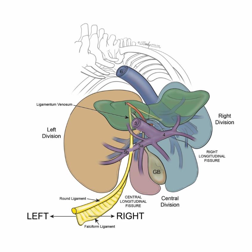
Portosystemic shunts are abnormal blood vessels that cause blood to bypass the liver, allowing toxins to build up. The shunts can develop in utero and lead to seizures, digestive problems, and stunted growth in puppies. Illustration: D. Sawchyn, MSMI, DVM, CMI of Sawchyn Medical
Illustration, LLC (SMI)
Cornell veterinary radiologists have discovered anatomical details about a birth defect that commonly affects liver function in dogs – a finding that could aid in life-saving surgical repairs for many canine patients.
Portosystemic shunts are abnormal blood vessels that cause blood to bypass the liver, allowing toxins to build up. The shunts can develop in utero and lead to seizures, digestive problems, and stunted growth in puppies.
Radiology resident Nicholas Walsh, D.V.M.'19 worked with Peter V. Scrivani ’89, D.V.M. ’93, professor of radiology, to investigate the anatomical components of this defect. They published the results this month in the journal of Veterinary Radiology and Ultrasound.
“Through our study, we developed a concise and accurate way to describe these birth defects that clarifies our overall understanding of their anatomy,” Walsh explained. “Portosystemic shunts are a complex topic where anatomy plays a big role in our treatment planning. Our discoveries are exciting because they have practical clinical applications for patients.”
An astonishing recognition
The imaging team evaluated CT scans of this birth defect in 56 dogs from Cornell’s Companion Animal Hospital and the Schwarzman Animal Medical Center in New York City. The scans zero in specifically on intrahepatic portosystemic shunts – abnormal blood vessels often described as going through liver tissue, instead of around it. The study revealed that about half of these abnormal vessels are actually sandwiched between liver lobes, but on the outside of the liver.
“For a lot of us who have been performing CT for a long time, this was an astonishing recognition,” Scrivani said. “This new way of characterizing portosystemic shunts will help us provide really good diagnoses and communicate better with surgeons to make the most of novel surgical treatments for this defect.”
For his work on the paper, Walsh earned the Best Scientific Content award at the 18th Cornell University College of Veterinary Medicine Clinical Investigators’ Day, an opportunity for residents and interns to present their research to the veterinary community and practice their oral presentation skills.
Full circle
Cornell alumni and current faculty members collaborated on the study. Lauren D. Sawchyn, D.V.M. ’09, a board-certified veterinarian and medical illustrator, created the diagrams in the paper. Anthony Fischetti, D.V.M ‘01, head of radiology at the Schwarzman Animal Medical Center, contributed to the paper. And veterinarians from Cornell’s Department of Clinical Sciences and the Department of Biomedical Sciences helped the team to understand the anatomy of this defect.
“It was such a collaborative effort,” Walsh said. “This is why I love radiology. We are here to work with and support so many of the various disciplines in veterinary medicine.”
For Scrivani, who recently was promoted to full professor Cornell, this paper brings his career full circle. In 1993, he was a senior veterinary student at Cornell when he first learned about portosystemic shunt imaging.
“I did my senior seminar paper on the imaging of portosystemic shunts,” he said. “Back then, we were limited in the imaging technology available. Today, we have advanced CT angiograms that give us a very detailed pictures of the liver and allow us to see the blood vessels in excellent detail.”
Scrivani hopes the discoveries in this paper will improve the care for canine patients across the globe. “We’ve already gotten emails from people around the world who are excited to use our new classification system,” he said. “We’re hopeful it will make a difference for patients.”
Written by Sheri Hall