Lyme disease in horses
This is an accompanying article to a Cornell Equine Seminar presented on Oct. 13, 2020 by Dr. Thomas Divers, the Rudolph J. and Katharine L. Steffen Professor emeritus of Veterinary Medicine in the section of large animal medicine.
Many horses are infected with Borrelia burgdorferi, the organism that causes Lyme disease – though clinical signs appear to be uncommon. Board-certified internist and emergency and critical care specialist Dr. Thomas Divers explains how Borrelia spreads, what clinical disease looks like, how it is diagnosed and treated, and what preventive measures you can take.
What follows here is an overview. Please watch Divers’s entire presentation for more extensive information.
Borrelia burgdorferi infection and Lyme disease
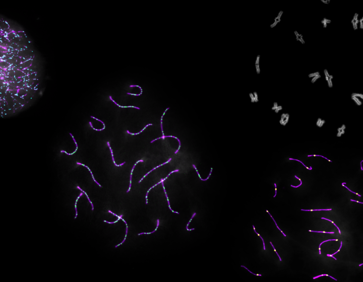
Lyme disease is caused by Borrelia burgdorferi, a tiny, coil-shaped organism (spirochete). It can infect many different species, including humans, horses, and dogs. White-footed mice and other small animals are reservoirs, and ticks become infected with B. burgdorferi when they feed on them.
The only tick that can carry and spread the organism to infect mammals is the blacklegged tick, also known as the deer tick or Ixodes tick. It is recognizable by a unique characteristic: “If you flip an engorged female Ixodes over, you can see the identifying anal port that has what some people call a McDonald’s arch all the way around it,” Divers explains.
It takes two years for the tick to mature from an egg to an adult. Larvae that have been exposed to the spirochete while feeding on rodents may infect humans. But adult ticks – and particularly females – are primarily responsible for spreading disease to horses and other mammals.
Once the tick begins to feed, Borrelia initiates both up and down regulation of certain genes and outer surface proteins (Osps) that help with transmission and survival of the organism. Antibodies against these proteins can be measured in a blood test and provide useful information. The presence of antibodies against OspC indicates recent infection within the last two to five months, while antibodies against OspF suggest a chronic infection. “If the horse has clinical disease, it’s much easier to treat in the early stages than in the chronic stages,” Divers says.
Antibodies against the OspA antigen were thought to be mostly caused by vaccination, but research showed that OspA antibodies are detectable in a relatively small percentage of naturally infected horses. Finding persistently high OspA antibodies in a non-vaccinated horse might be more predictable of clinical disease than are antibodies against C and F. “More research is needed on the relationship of clinical disease with high OspA levels,” Divers says.
Since testing for antibodies, so-called seroprevalence studies, began around the year 2000, there has been strong evidence that the geographic range of blacklegged ticks and Borrelia burgdorferi exposure in animals have been increasing in North America. Exposure and infection are especially high in horses because they readily have contact with ticks. “But clinical disease in horses that are infected appears to be relatively rare,” Divers explains. It is also unclear whether chronic infection, shown predominantly by the presence of antibodies against the proteins OspF or C6, could become activated into clinical disease at some point.
“There’s a lot we don’t know about equine Lyme disease,” Divers says. He recommends a test only if a veterinarian suspects clinical disease and discourages routine testing of healthy animals.
What happens with experimental infection
A study conducted at Cornell traced what happens when ponies are infected with Borrelia. Most showed no clinical signs but had some pathology, leaving open the potential for clinical disease to develop. “We also learned where the organism tracks in the equine body,” Divers says.
The tick needs to be attached for at least 18 hours for the spirochetes to undergo the sophisticated process of upregulating and downregulating specific antigens that will allow them to enter and survive in the horse. They then migrate mostly through the skin and connective tissue to joint and synovial membranes. Some also go to the covering of the brain and the spinal cord (the meninges) and to the eye.
Clinical signs that may show up due to this movement are swollen lymph nodes near the site of the tick bite, dermatitis, hyperesthesia – increased sensitivity in the horse when it is touched – stiffness, multiple limb lameness, inflammation in the joint capsules, and occasionally meningitis or uveitis.
Clinical disease in horses
The best documented clinical disease syndromes in horses with Lyme disease are:
- neuroborreliosis, the neurologic form of meningitis
- uveitis, inflammation of the uveal tissue in the eye
- cutaneous pseudolymphoma, nodules around the skin, often at the site of the tick bite, that can easily be treated with antibiotics
- bursitis
Many undocumented clinical signs seen by veterinarians and owners include:
- stiffness, often in all four legs
- behavioral changes related to nerve irritation
- shifting leg or intermittent lameness
- muscle wasting due to inflammation in the nerves supplying the muscles of the neck or back
- hyperesthesia
“These haven’t been proven, but I have no doubt they do occur,” Divers says. “It’s going to take a lot more work to sort out all the different ramifications of Lyme disease in horses.”
Lyme uveitis
Several cases have been reported in which horses had unusual uveitis and proved to carry Borrelia in their eyes, primarily in the vitreous fluid. These cases usually progress very fast and in both eyes, sometimes turning horses blind within two weeks. Lyme uveitis may precede the onset of central nervous system signs.
Clinical signs reported with neuroborreliosis
When the Lyme organism affects nervous tissue, mostly around the spinal cord and sometimes in the brain or peripheral nerves, horses may show these signs:
- muscle atrophy
- weight loss
- cranial nerve deficits, e.g. the inability to swallow properly, facial paralysis
- ataxia and paresis, i.e. coordination issues when the spinal cord is involved
- change in behavior, including narcolepsy
- hyperesthesia
- fasciculations, i.e. muscle twitching
- neck and back stiffness, where some horses will not lift their head above a horizontal plane
Diagnosing Lyme disease
The first step in diagnosing Lyme disease is testing for antibodies, though most horses will show this evidence of exposure.
Antibodies need to be paired with clinical signs that are compatible with Lyme disease in horses or other species, such as hyperesthesia or pseudolymphoma. “Make sure that other diseases that could cause those clinical signs are ruled out before jumping on the Lyme disease bandwagon,” Divers says.
The diagnosis of Lyme disease is supported by the microscopic findings of lymphohistiocytic and plasmacytic (immune cell) infiltrate in diseased tissue. Another, more modest, piece of evidence is thickening of the dura – the covering of the spinal cord and brain – due to chronic inflammation. “Very few equine diseases will do this,” Divers explains.
 Which horses should we treat?
Because a positive antibody test does not predict future clinical disease, Divers recommends not treating based on antibodies alone, which will allow you to avoid unnecessary expense, an increased risk of issues with the medication, and inappropriate use of antimicrobials.
Only horses with antibodies and clinical signs consistent with Lyme disease, for which other potential causes have been ruled out, should be treated. Horses with elevated OspC could be an exception, because it indicates recent infection that is easier to treat at this stage, but Divers still considers the risk of developing clinical disease low and is not a proponent of treatment based upon elevated OspC antibodies without clinical signs. (Divers notes that some confirmed cases of Lyme disease are not positive serologically for B. burgdorferi, and virtually all of these cases have been documented to have an immune deficiency.)
Treatment of equine Lyme disease
Treatment recommendations for equine Lyme disease have been based on lab studies, studies done in humans and dogs, organ system involvement, available data on bioavailability of drugs and their half-life and distribution in the body (pharmacokinetics) and their safety and cost. The only experimental treatment trial in equines was performed at Cornell in 2000.
There is no consensus on a preferred drug for treating equine Lyme disease, but in the pony study intravenously administered tetracycline cleared the organism completely, while some ponies that received orally administered doxycycline or intramuscular administered ceftiofur remained infected. Beta lactams – drugs such as penicillin, ampicillin, and other cephalosporins – are also potentially effective.
Although there is an established “standard of care” treatment duration for human Lyme disease, it does not apply to horses because the infection in horses may be present for a lot longer than in humans before disease detection. Drugs such as doxycycline or minocycline are also significantly less bioavailable in horses, so higher doses have to be given at greater expense. Oral penicillins and most beta lactam antibiotics are not recommended to be given orally in horses because of poor bioavailability and high risk of gastrointestinal upset. “So it’s much more difficult to treat Lyme disease in horses than in people or dogs,” Divers says.
Ideally, once antibiotic treatment has begun, antibodies should decline continually over a three to four-month period down to what are considered negative levels.
Ancillary treatments
Divers does not recommend giving corticosteroids, which suppress the immune response, except if the infection is in the eye or the central nervous system. “These are critical, unforgiving organs,” Divers says. Non-steroidal anti-inflammatory drugs (NSAIDs) may help the horse feel better but otherwise have minimal benefit. Finally, Divers and colleagues scoured both the veterinary and human literature for studies on acupuncture and herbal treatments but could not find any high-quality scientific evidence that they are effective.
Prognosis
“In horses with uveitis and neurological disease, the prognosis is poor, it’s as simple as that,” Divers says. In other cases, horses frequently improve and go back to normal. If the organism hasn’t been completely cleared, they may relapse with almost identical clinical signs, so a longer antimicrobial treatment may be warranted.
Tick control and vaccination
For Divers, tick control is at the heart of prevention. Adult female Ixodes ticks are thought to be responsible for the highest percentage of B. burgdorferi horse infections. They predominantly feed in early spring or late fall, putting horses at the greatest risk during these times. With global warming, infections can now occur in the winter months in the northeastern United States.
The few available treatments for tick control include pour-ons and sprays with permethrin, but they are relatively short-acting. More research is needed on tick control in horses, possibly using some canine approved products. Divers suggests keeping pastures clipped so ticks can’t crawl up tall grasses and keeping horses away from brush areas on the edge of fields. After bringing horses in from the field or after trail riding, a close inspection of the horse for ticks should be performed and any tick found should be properly removed.
A vaccine using OspA – a protein that Borrelia normally downregulates during natural infection was shown in a study to produce a good immune response in horses. It and other surface-based antigen or whole cell canine vaccines are available, but in horses the high levels of vaccine-produced antibodies don’t last long, so the horse would need to receive a series of boosters. “You may want to time your first vaccinations and your booster so that you have good antibody levels in the spring and the fall,” Divers advises. He believes that a hypothetical vaccine based on OspA and OspC together could be more effective, but OspA-based vaccines have been used the most.
Equine and human Lyme disease: Controversies
Lyme disease in humans and horses shares many similarities, from the infecting organism to the means of transmission and diagnostic tests used – though Divers points out that some veterinary tests are simpler and faster than those in human medicine. But none of the tests pick up the antibody early after infection.
In both species, recurrent or persistent signs and continued positive antibody tests are generally considered inadequate to confirm a diagnosis of chronic Lyme – nor are improvements in symptoms after a horse is put on antibiotics, as the tetracycline family of antibiotics can have general anti-inflammatory properties, Divers explains. Nevertheless, both humans and horses are often put on long courses of antibiotics without good cause. The key, Divers emphasizes, lies in adequate tick control foremost and working towards an approved Lyme vaccine with a long duration of immune protection.
Future directions
Divers believes Lyme disease is likely overdiagnosed. “We need better ways to prove not just exposure but detection of the organism in the body of the patient,” he says. Studies should also focus on identifying the range of clinical signs associated with the disease. In addition, Ixodes ticks carry other pathogens whose effects in horses have not yet been widely studied:
- B. mayonii is currently reported only present in Wisconsin and Minnesota and cross-reacts with B. burgdorferi tests, turning them positive.
- B. miyamotoi is present in the Northeast and other areas of the U.S. and does not serologically cross-react with B. burgdorferi. While it has been reported to cause disease in humans, it has not yet been proven to infect or cause disease in horses.
- Powassan virus has been found in New York State in the past few years during the summer. In humans, it produces high fevers and occasional encephalitis. Transmission occurs quickly following tick attachment. More studies are needed on transmission and possible disease in horses.
Lyme and anaplasmosis
Many ticks are infected with both B. burgdorferi and the bacterium Anaplasma phagocytophilium, which causes anaplasmosis. Well-documented in horses, disease occurs almost solely in adult horses. Their symptoms mostly include high fever, depression, leg edema, jaundice, and low white blood cell and platelet counts. Other signs may occasionally be found, and in some horses the infection causes no noticeable disease.
Within five or six days of infection, the organism shows up in the white blood cells, unlike Borrelia, which appears in connective tissue, skin, and nerves, and rarely in the blood. The high fever is another difference between the diseases, as most horses with Lyme disease are afebrile.
In contrast to people, who when infected with A. phagocytophilium can sometimes develop acute multiple-organ system failure and septic shock, horses have low mortality from anaplasmosis. Untreated, they will usually get better within 10 days. To shorten their suffering, Divers recommends a couple of doses of oxytetracycline – IV-administered works best – and a one-week course of minocycline or doxycycline.
While Lyme and anaplasmosis were rare in central New York when Divers first came to Cornell in 1990, they are now much more common. “It’s the ticks,” Divers sums up. “The ticks are moving, and they take their infectious agents with them.”
Watch Divers’s full talk online, and see the entire Cornell Equine Seminar Series videos here.
Addendum: Dr. Divers would like to recognize the early work on equine Lyme disease by Dr. Yung Fu Chang at Cornell University. “Dr. Chang’s early research on B. burgdorferi infection studies in ponies, treatment trials and recombinant OspA Lyme vaccine studies have provided the foundation for what we know about Lyme disease in horses,” he says.
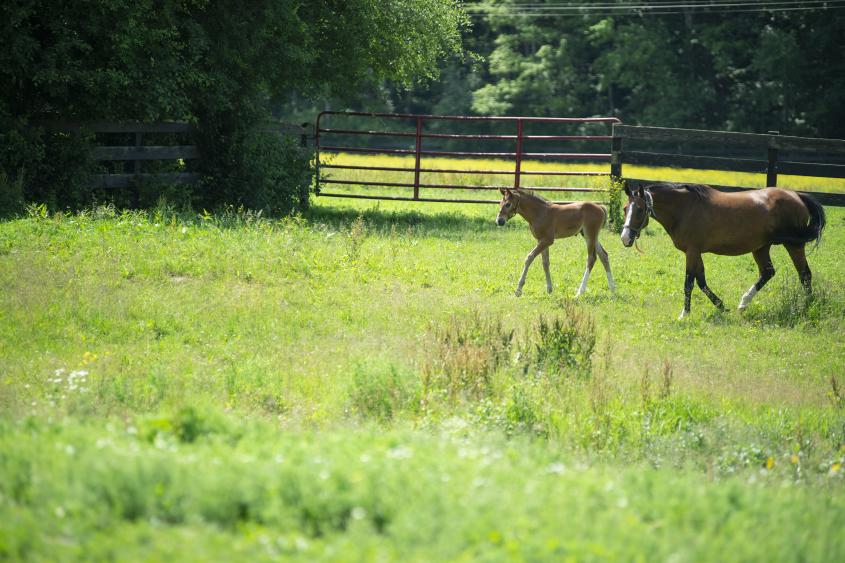
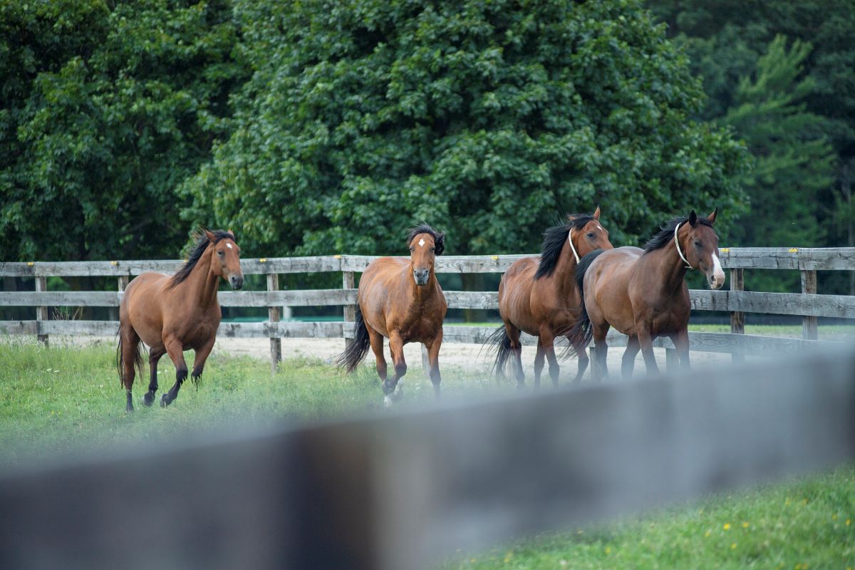
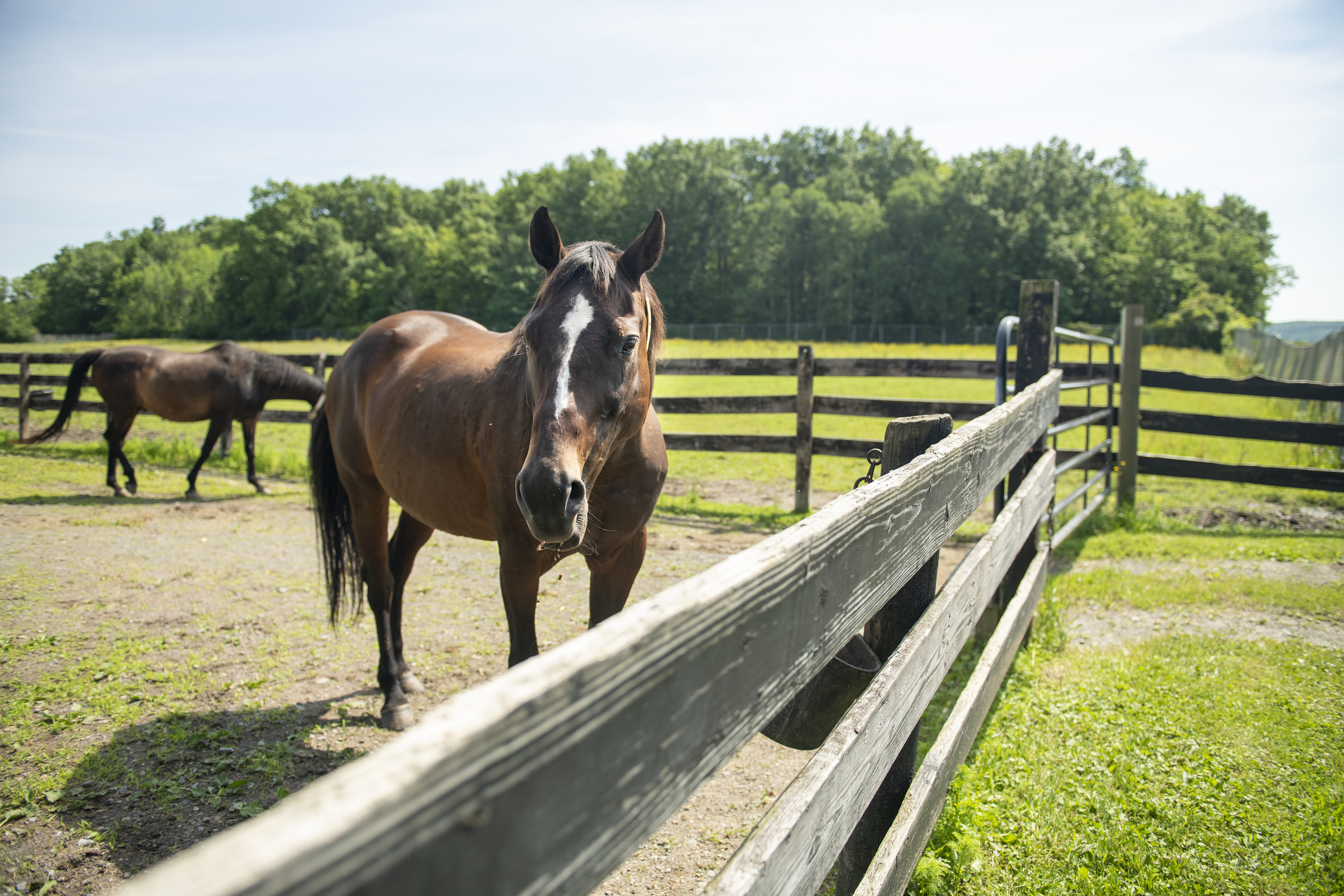
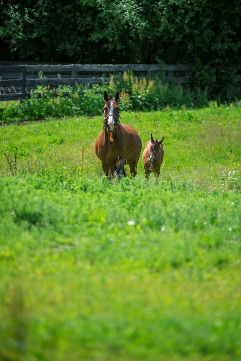
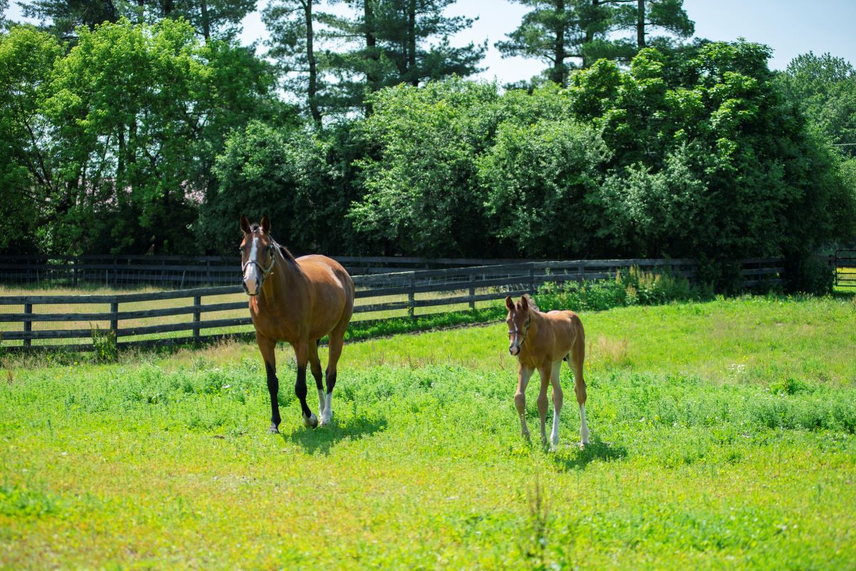
Photos by Rachel Philipson