


Illustrated tools for improving canine nictitans gland prolapse surgical repair skills
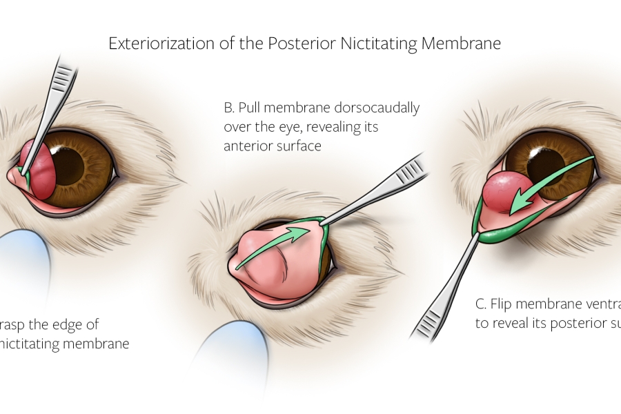
The surgical repair of a prolapsed nictitating membrane (NM) gland, also called cherry eye, is a common issue encountered in veterinary clinical practice. For successful surgery, constant repositioning of the NM gland is necessary, but the technique is difficult to conceptualize and understand. To help break down this complex process into easy-to-follow steps, ESS’s Medical Illustrator Allie Buck, in collaboration with CVM’s Dr. Eric Ledbetter, created a series of surgical illustrations to show the necessary steps for executing correct surgical technique. Students learn to appreciate the impact that successive repositioning of the nictitating membrane has on local anatomy.
Project Support
This project was made possible by a Veterinary Curriculum Enhancement Grant (2020 cohort) from the Cornell University College of Veterinary Medicine.