


Cherry eye in dogs
Overview
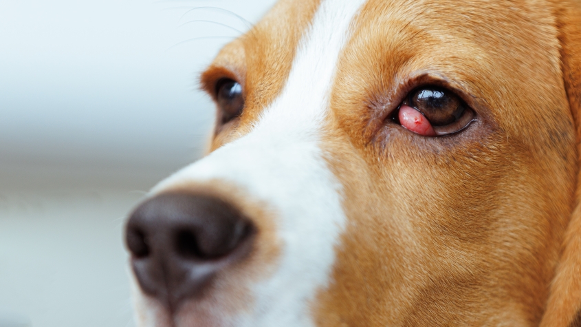
“Cherry eye” is the common name for a prolapse of the third eyelid gland in dogs. The third eyelid is a thin membrane located at the inner corner of a dog’s eye. It contains an important tear-producing gland that normally is not outwardly visible. When this gland pops out from its normal position, it appears as a smooth, pink or red mass at the corner of the eye, resembling a cherry.
Because this gland produces a large portion of the eye’s tears, a prolapse can interfere with normal tear production. This may cause eye redness, discharge and irritation. The treatment for cherry eye is surgical replacement of the gland.
Cause
The exact cause of cherry eye is not entirely known, but genetics are thought to play a role as certain dog breeds appear predisposed, including American and English Cocker Spaniels, Beagles, English and French Bulldogs, Pugs and Boston Terriers. Cherry eye is much more common in younger dogs, typically under two years of age, and can affect one or both eyes.
The third eyelid, also called the nictitans membrane, contains a T-shaped piece of cartilage and one of the most important tear glands (third eyelid gland), which produces up to half of all the watery portion of the tear film. The tear film keeps the eye lubricated and healthy and is distributed across the eye’s surface by the third eyelid and eyelids. When the gland prolapses, it can become inflamed, irritated or infected, and tear production may be reduced.
A different condition, called scrolled cartilage, can look similar to cherry eye. In this condition, a portion of the cartilage of the third eyelid grows abnormally and bends or “scrolls,” creating a pink or red swelling in the same location. Scrolled cartilage is less common but tends to occur more often in giant breeds such as Great Danes. Scrolled cartilage can occur independently or concurrently with cherry eye, and in some cases, the two conditions can only be distinguished during an exam under anesthesia.
Clinical signs
- A smooth, round, pink or red mass in the inner corner of the eye
- Eye discharge
- Eye redness
Diagnosis
Cherry eye is diagnosed on a physical exam based on the appearance of a pink or red, smooth mass of tissue protruding from the inner corner of the lower eyelid. A test to evaluate tear production (Schirmer tear test) may be performed. The third eyelid will also be evaluated for scrolled or deformed cartilage and, in rare cases, more serious conditions such as cancer.
Treatment
The treatment for cherry eye is surgical replacement of the gland. Your veterinarian may perform the procedure or refer you to a veterinary ophthalmologist. Several surgical techniques exist, and the best option depends on your dog’s needs and the surgeon’s preference.
Surgical removal of the gland is not recommended because dogs that lose this gland are at high risk of developing chronic dry eye (keratoconjunctivitis sicca, or KCS). KCS is a lifelong condition that requires daily medications to keep the eyes comfortable.
If scrolled cartilage is present, surgery may include trimming, or otherwise reshaping, the abnormal portion of cartilage.
After surgery, your dog may need short-term eye medications to prevent infection and manage pain and inflammation.
Outcome
The prognosis for dogs after surgical correction of cherry eye is generally very good. Replacing the gland soon after it prolapses often gives the best chance of restoring normal function. Without surgery, dogs are at significant risk of ongoing inflammation and developing chronic dry eye (KCS), an uncomfortable condition that requires lifelong topical eye medications.
Recurrence is possible after surgery, especially if the prolapse has been present for a long time, if the gland is significantly inflamed, or if abnormal cartilage of the third eyelid is not corrected. The success of the procedure may vary based on the surgical technique, surgeon, and individual patient.
This page was last updated on Tuesday, Dec 02, 2025