Imaging Service

The Imaging Service at the Cornell University Hospital for Animals takes pride in providing excellent patient care and customer service.
Medical imaging is the process of creating and interpreting images of a body’s morphology and function for the explicit purposes of patient examination and intervention. It also is a subset of biological imaging, which refers to the use of some imaging technologies that noninvasively capture information about the deep internal morphology and function of a patient.
In veterinary medicine, some of the most commonly used imaging technologies are radiography, ultrasonography (US), computed tomography (CT), and magnetic resonance imaging (MRI). As a clinical specialty, medical imaging has broad impact on patient care and is used by a wide range of health care professionals in addition to radiologists. The broad clinical impact has led to the development of many subspecialties, which are based on patient management goals, imaging technologies, body parts or systems or patient characteristics. Some radiologists perform radiation therapy. At Cornell, the Imaging Service performs radioiodine therapy for feline hyperthyroidism.
We maintain the most current technology and employ an experienced staff to obtain medical images and diagnose routine and complex diseases in large, small and exotic animals. Our licensed technicians, board-certified radiologists and radiology residents-in-training perform and interpret imaging examinations on a wide range of species. Our team of veterinary imaging experts are leaders in clinical research, education, resident training and clinical practice. We strive to discover, preserve and share knowledge about veterinary imaging on local and global levels.
What to Expect During Imaging
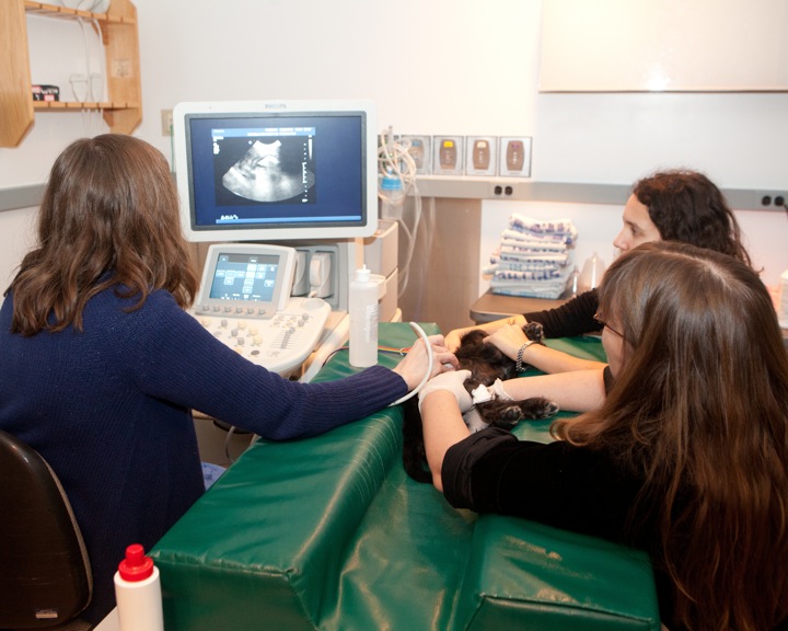 If you have an appointment for an outpatient imaging examination, check in at the main reception desk. After registration, a technician will meet you in the reception area, answer questions and take your pet for their examination. For most ultrasound examinations, you may accompany your pet.
Your animal may need to be sedated or anesthetized to prevent movement during imaging. We do this when we anticipate that the procedure might be uncomfortable or painful, if the animal is anxious and whenever we expect that not holding perfectly still would defeat the procedure – motion is the bane of imaging! We work with your primary Cornell veterinarian to decide which sedative or anesthetic is most appropriate. Radiographs and ultrasound are often done awake, sometimes with a sedative and occasionally, when necessary, we manually restrain the animal. We minimize manually holding animals for radiographs to limit our own exposure to radiation. Animals receiving a CT are sedated or anesthetized but MRI patients are almost always given general anesthesia. Anesthesiologists are available to consult on all cases and ensure the safety of your animal.
We interpret images and provide an oral report on the same day the study is obtained followed by a written report. For hospitalized animals, emergency imaging and interpretation is available 24/7/365 by residents and faculty on back-up.
Imaging Modalities
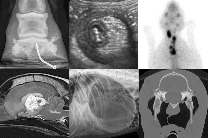
Radiography
Agfa DX-G CR
Commonly referred to as “x-rays”, radiographs are used widely in all species for examination of any body region. Radiographs are often the initial modality used to investigate a disease problem and can be diagnostic on their own or useful when recommending additional types of imaging.
Ultrasound
Philips EPIQ5 and Philips IU-22
Sonography is commonly used by radiologists to evaluate the internal structure of organs, search for fluid accumulations, evaluate blood flow, detect infections, tumors, and help determine the extent of trauma. While abdominal sonography is our most common examination, all body regions can be examined. Our cardiologists use ultrasound for echocardiography – a specialized examination of the heart.
Computed Tomography (CT)
Toshiba Aquilion Large-Bore, 16-slice
CT scanning uses x-ray and sophisticated computer processing to render cross-sectional images of the body. The current CT equipment is fast enough to scan through an entire large-breed dog in under a minute and much less for a cat, rabbit or other small animal. While general anesthesia is necessary for some examinations, others can be obtained with sedation or very short acting anesthetics leading to faster and safer examinations. CT scanning is useful in the evaluation of trauma, neurological conditions in the back, chest and abdominal cancers, head and neck diseases, and for planning both surgical and radiation cancer treatments.
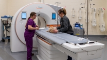
Magnetic resonance imaging (MRI)
Toshiba Vantage Atlas 1.5 Tesla
MRI examines body structures using the property of nuclear magnetic resonance to stimulate with radiofrequency energy the nuclei of hydrogen atoms aligned in a strong magnetic field and detect those signals for imaging. MRI provides excellent contrast between soft tissues that are otherwise indistinguishable by other means, which makes it especially useful in imaging the nervous (brain and spinal cord) and musculoskeletal (muscles, tendons, ligaments) systems.
Nuclear Medicine
MIE Equine Scanner HR/Scintron VI-VME
In nuclear medicine imaging, radiopharmaceuticals are injected into the animal and a gamma camera detects the distribution of the isotope in the body. These examinations are used mainly for the diagnosis of bone, thyroid, kidney and liver diseases. The radiation dose from the isotope is minimal and use is fully regulated by Environmental Health and Safety at Cornell and the New York State Health Department.
We offer I131 Radioiodine treatment for cats with hyperthyroidism.
Digital Fluoroscopy
Philips R/F and Ziehm C-arm
Fluoroscopy guides interventional procedures (placement of pacemakers, occlusive coils, and catheters) and is used to examine organ function (swallowing, tracheal collapse syndromes, and gastrointestinal motility). Examinations may be performed in the radiology suite on awake animals. The “C-arm” systems permit fluoroscopy in the operating room during surgery for real-time guidance in catheter placements and orthopedic repairs and prosthetic implantations.
Related Information
American College of Veterinary Radiology
The ACVR sets standards for veterinary imaging professionals, certifies training programs, and examines residents during and at the completion of a three-year, post-graduate program. Passing a written and oral examination leads to “board-certification” and is the credential required to practice as a veterinary radiologist. CUHA radiologists are board-certified and dedicated to service, education and research in veterinary imaging. We regularly have three or four residents training in the Hospital.
Veterinary Diagnostic Imaging Residency Program
The American College of Veterinary Radiology (ACVR) accredits Cornell’s residency program in Veterinary Imaging. This 4-year program generally enrolls one new candidate each July and provides specialty training in radiology, fluoroscopy, ultrasonography, nuclear medicine, computed tomography, and magnetic resonance imaging in small and large animals, and radioiodine treatment of hyperthyroidism in cats.