


Research
Comparative Mammalian Reproductive Endocrinology/Physiology and Aging
My lab's principal area of interest is female reproductive physiology and aging in mammals, with a focus on ovarian function. We perform integrative studies at the molecular, cellular, and organismal levels to elucidate the mechanisms that underlie and potentially attenuate reproductive aging.
My lab generally follows Krogh’s Principle: “For a large number of problems, there will be some animal of choice, or a few such animals, in which it can most conveniently be studied.” (1929).
Two questions that are presently being addressed with interesting animal models are:
1. How do long-lived female mammals maintain and establish an ovarian reserve that will last for the duration of their reproductive lives?
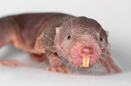
To address this question, we are studying naked mole-rats (NMRs, Heterocephalus glaber), because they are the longest lived rodent and females demonstrate no decline in fertility and fecundity into their third decade of life. This work is in collaboration with Dr. Melissa Holmes (University of Toronto, Mississauga) who maintains an extensive NMR colony system for her studies on their neurobiology. NMRs are comparable in size to lab mice, and in collaboration with Dr. Miguel Brieño-Enriquez, we have found NMRs have an unusual abundance of oocytes (eggs) and that the process leading to the establishment of the ovarian reserve is developmentally delayed. We are also evaluating a transgenic mouse line generated by Dr. Vera Gorbunova's lab (University of Rochester) that expresses the NMR gene for hyaluronan synthase 2 (nmrHas2) to determine whether female reproductive aging is attenuated in nmrHas2 mice. In collaboration with Drs. Francesca Duncan (Northwestern University) and Michele Pritchard (University of Kansas Medical Center), we are investigating the impact that the NMR's very high molecular weight hyaluronan has on ovarian inflammation and fibrosis as females age.
2. Can anti-Müllerian hormone (AMH) be used to optimize exotic animal breeding programs?
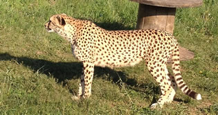
To address this question, we are studying cheetahs (Acinonyx jubatus), because ex situ populations of these big cats carry a disproportionate number of females that are beyond their prime reproductive age. This work is in collaboration with Drs. Adrienne Crosier (Smithsonian Conservation Biology Institute). AMH correlates with the size of the ovarian reserve in other mammals, and our preliminary findings suggest female cheetahs follow this aging-related pattern. However, cheetahs of the same chronological age can have markedly different AMH concentrations. We are in the process of determining if AMH helps to predict best outcomes for ovarian stimulation protocols when they’re used for assisted reproductive technologies, e.g., IVF.
Methods used:
- Ovarian histology and immunohistochemistry
- Hormone assays
- Quantitative real-time PCR
- single cell/nucleus RNA-seq
- Transgenic mice
Animal models previously investigated:
Spotted hyenas – Natural masculinization of female external genitalia
Siberian hamsters – Short day lengths delayed female reproductive aging
Syrian hamsters – Chronological, but not reproductive age, influenced female mate preference
Yellow-pine chipmunks – Hormones and the hormonal response to stressors varied across seasons