


Imaging
Imaging Service
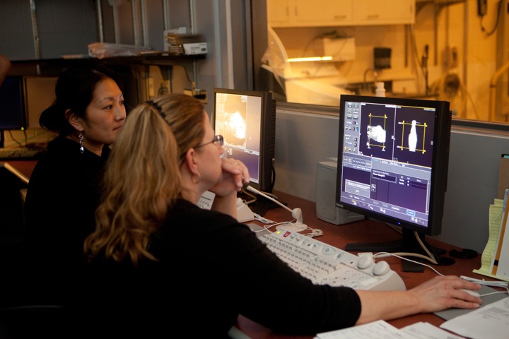 The Imaging Service at the Cornell University Hospital for Animals takes pride in providing excellent patient care and customer service.
Imaging Service

The Imaging Service at the Cornell University Hospital for Animals takes pride in providing excellent patient care and customer service.