


Ophthalmology
Ophthalmology
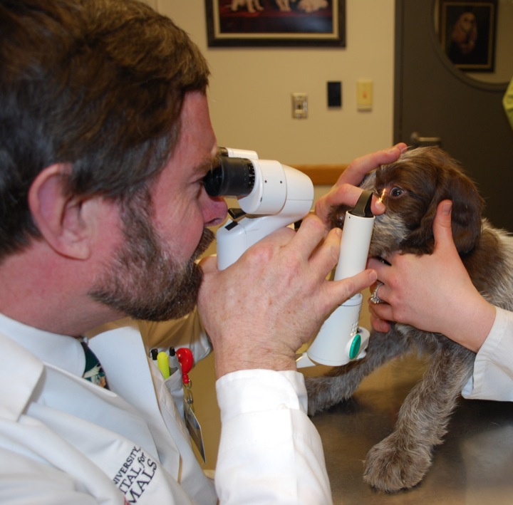 The Ophthalmology Service at the Cornell University Hospital for Animals provides scheduled and emergency care for companion animals with eye and vision problems. Our staff include the board-certified ophthalmologists and residents who collaborate with other veterinarians across the Northeast to provide comprehensive eye care.