


Equine Enteric Coronavirus
The Cornell University Animal Health Diagnostic Center (AHDC) has seen a rise in the diagnosis of equine enteric coronavirus cases since initial outbreaks were investigated starting in 2010. The AHDC Veterinary Support Services veterinarians are attributing this increase in equine enteric coronavirus in our area of the Northeast to improved awareness of the disease and therefore diagnostic submissions. Since 2013, nearly 2000 samples have been submitted to the AHDC for equine coronavirus testing, of which approximately 18% have tested positive.
Equine Enteric Coronavirus
Overview
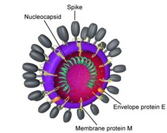
Coronaviruses comprise a large group of RNA viruses that can cause both respiratory and enteric signs of disease in various species. They are further grouped based on genetic and serologic differences into alpha, beta and gamma coronaviruses. The equine coronavirus, a beta coronavirus, has been recently isolated from a number of outbreaks across the United States, Europe and Japan, with its rising incidence being associated with increased awareness and testing. Equine coronavirus manifests as an enteric disease in the horse. Seroprevalence of equine coronavirus in the Unites States has been estimated at 9.3% (Kooijman et al., 2017).
Transmission
Transmission of equine coronavirus is via the fecal-oral route. Although beta coronavirus commonly causes enteric and respiratory symptoms in cattle, the prevalence of equine coronavirus in the nasal secretions of horses with fever and respiratory disease is low (Pusterla et al. 2015). This finding may suggest a lack of tropism by equine coronavirus for the equine respiratory tract epithelium.
Incubation Period
Clinical signs of equine coronavirus are seen 48-72 hours after exposure and fecal viral shedding begins 3-4 days after exposure. Peek shedding is documented 3-4 days after the development of clinical signs (Pusterla et al, 2018). This timeline of shedding may cause horses to test fecal PCR negative during the very early stages of clinical disease.
Duration
Clinical signs generally resolve in several days to 1 week with supportive care and outbreaks typically last for about 3 weeks (Pusterla et al., 2013). Fecal viral shedding has been documented to more commonly range from 3-25 days, however cases have been documented to shed as long as 99 days (Goodrich et al. 2018) and anecdotal reports suggest intermittent shedding may occur. Asymptomatic shedders do exist and may be a source in the spread of disease (Pusterla et al, 2018). These horses will not show clinical signs but are in fact shedding the organism in their manure.
Survival in the environment is unknown. Although a direct comparison cannot be made, human coronaviruses have been shown to survive longest at cooler temperatures (39.2F) for 14 days in waste water and 17 days in feces.
Age Distribution
Equine coronavirus is seen as a mono-infection in adults, usually older than 2 years of age. When seen in foals, equine coronavirus has been documented as a co-infection with Rotavirus or Clostridium perfringens (Pusterla et al., 2018).
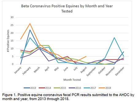
Seasonality
More commonly seen during the cold weather months (in the Northeast areas), October through April, but cases are also diagnosed in the heat of the summer.
Common Clinical Signs and Bloodwork Abnormalities
- Anorexia
- Lethargy
- Fever (101.5-106F)
- Changes in fecal character (soft formed); in severe cases profuse diarrhea can occur but is not routinely seen
- Mild colicy-like signs (laying down; looking at sides)
- Leukopenia secondary to neutropenia or lymphopenia. The hemogram can be unremarkable.
- Hypoalbunemia
- Neurologic abnormalities (ataxia, depression, recumbency) secondary to hyperammonemia. Hyperammonemia-associated encephalopathy can be seen in severe cases secondary to increased ammonia production by the overgrowth of urease-producing bacteria or secondary to increased ammonia absorption through the disrupted intestinal mucosal barrier.
Morbidity
Morbidity ranges from about 10-83% and death is typically rare. When death occurs it is thought to be secondary to complications associated with gastrointestinal barrier disruption leading to septicemia, endotoxemia and hyperammonemia-associated encephalopathy (Pusterla et al., 2018).
Case Management
Treatment most often is supportive, which may include fluid therapy and non-steroidal anti-inflammatories. More severe cases may require extensive treatment or hospitalization.
AHDC Sample Submission/Requirements
The sample for equine coronavirus testing is fresh feces submitted in an unbreakable leak-proof container to the laboratory by overnight courier on ice packs. Samples must be kept chilled to prevent overgrowth of bacteria that may cause inhibition in the PCR testing. If sample submission may delayed for more than 3-4 days, feces should be frozen to prevent bacterial overgrowth. Parasitology cannot be performed on frozen fecal specimens; therefore, if both tests are to be requested both fresh and frozen feces must be submitted. Feces are tested by: Equine Enteric Corona PCR. Lag Time: 3 business days. For any questions contact the AHDC and ask to speak to the Veterinary Support Services (VSS) team at 607.253.3900.
Biosecurity/Control Measures
- If beta coronavirus is on your differential list, encourage the barn to practice appropriate biosecurity measures to control the spread of the virus.
- For biosecurity, see the AAEP guidelines.
- Horses can continue shedding the virus in their feces for weeks beyond the resolution of their clinical signs. Encourage the farm to take precautions by using footbaths, individual thermometers, and disposable gloves between horses. Attempt to isolate affected animals, handle them last and use separate manure handling equipment from the rest of the barn. Minimize traffic into/out of barn.
- Given the intermittent fecal shedding and published and anecdotal reports for shedding duration of equine coronavirus, it is difficult to make firm recommendations on testing after an illness or infection with the virus. It would be good practice to quarantine/isolate any new arrivals for approximately 21 days. This is in line with most recommendations for new entries into a new stable area.
- Disinfectants that are effective at inactivating equine coronavirus include sodium hypochlorite (bleach), povidone iodine, chlorhexidine gluconate, phenols, quarternary ammonium compounds and peroxygen compounds. It should be remembered that organic materials (feces, bedding, etc.) will decrease the efficacy of many disinfectants.
References
- Fielding C.L. et al. 2015. Disease associated with equine coronavirus infection and high case fatality rate. J Vet Intern Med. Vol 29. Pp 307-310.
- Giannitti F. et al. 2015. Necrotizing enteritis and hyperammonemic encephalopathy associated with equine coronavirus infection in equids. Veterinary Pathology. Vol 52(6). Pp1148-1156.
- Goodrich E. L. et al. 2018. Novel findings from a beta coronavirus outbreak on an American Miniature Horse breeding farm in upstate New York. Equine Veterinary Journal. Vol 12938. Pp 1-5.
- Kooijman L.J. et al. 2017. Seroprevalence and risk factors for infection with equine coronavirus in healthy horses in the USA. The Veterinary Journal. Vol 220. Pp 91-94. Pusterla N et al. 2018. Enteric Coronavirus infection in adult horses. The Veterinary Journal. Vol 231. Pp 13-18.
- Pusterla N et al. 2015. Prevalence of equine coronavirus in nasal secretions from horses with fever and upper respiratory tract infection. Veterinary Record. Sept 19.
If you have questions, please contact Veterinary Support Services.
VSS-WEB-092-V02 9/23