


A comprehensive online dentistry curriculum is now available. This program is an efficient and engaging way to deepen a learner's knowledge of one of the fastest-growing clinical disciplines in the field.
Taught by some of the world leaders in veterinary dentistry, our courses guide participants through the gold standard of dental care, using real cases that combine practical and conceptual knowledge.
Course details
This program is entirely online and self-paced. It can be run in facilitator- and/or faculty-led segments or as self-paced modules for participants to complete. Each of the three courses in this program offers comprehensive training and discussion on crucial components of veterinary dentistry. Participants will learn how to apply the gold standard of care using three diagnostic methods, which gives them the most accurate information for their patient’s health now and in the future.
Information for veterinary educators
Whether you are faculty looking to augment your students’ theoretical knowledge of dentistry or a curriculum designer seeking a new method for competency training, this program can be customized to meet the unique needs of your students.
Watch Dr. Nadine Fiani explain how adopting this program will unite the latest theory and gold-standard practices of veterinary dentistry into your curriculum:
Why adopt it?
For more information, download our helpful PDF here.
How does it work?
 As a curriculum designer for your veterinary school, you have the option to meet with our course leaders and faculty to see how you can most effectively incorporate this program into your curriculum. We offer custom guidance on its integration.
Most ideally, this program functions as a bridge between theoretical knowledge and practical application, and thus would fit best for third-year veterinary students just embarking into the clinical space.
You can use this program as self-directed learning for your students or incorporate it into an existing didactic course. It is easy to create a practical lab, either cadaver-based or with live patients, using the knowledge gained from these courses.
For groups of over 20 users, you will have access to a sign-in portal and dashboard custom-made for your group that is FERPA compliant. Through this portal, your students’ data stays together, is downloadable, and you’re able to easily assign admins who can view their progress and grades.
Moreover, if you purchase bulk access to this program, you’re entitled to an educational institution discount. You can also elect to have students purchase their own seats through your custom portal.
Each course comes with a final assessment that can be integrated into your curriculum, and the program as a whole can be used as a standalone course, integrated into an existing course, or augmented by hands-on labs.
What does this offer your students?
The overall aim of this program is to fill the gap between learning theory and jumping into practice. To that end, we’ve created practical material that takes veterinary dentistry theory and puts it into the context of what students will be faced with when working with actual patients. In doing so, they’ll engage with interactive 3D models, high-quality clinical photos, videos and more.
This program teaches
- The benefits of incorporating dental exams and intraoral radiology in daily clinical work
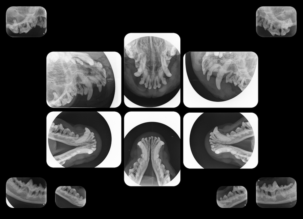
- The principles of intraoral radiography, and the correct use of radiology equipment and techniques to obtain and interpret diagnostic quality intraoral radiographs, including a full-mouth series
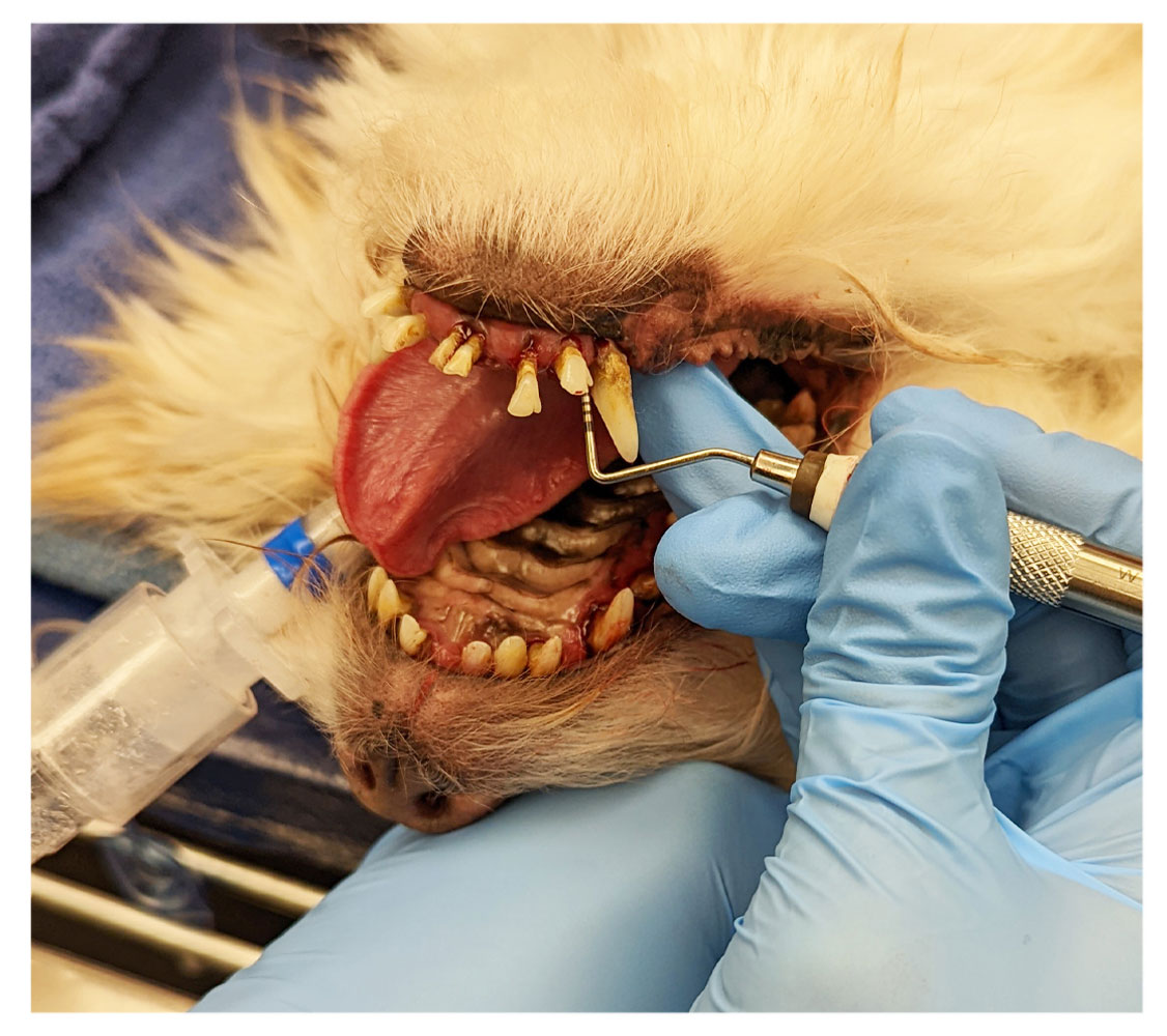
- How to perform conscious and anesthetized oral examinations, including how to compile and record significant findings and investigate abnormalities
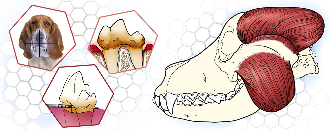
- The normal anatomy of dental structures as it pertains to common diseases
- How to create and maintain a safe environment for patients and clinicians, including clinician ergonomics
What does this offer your faculty?
This is a self-paced program, offering practical knowledge that can be applied either as a standalone, self-directed learning opportunity — or as an element of your existing dentistry course, all under your control.
We have put years of effort into creating this program so that you don’t have to. Faculty receive admin access to these courses, and each offers an assessment so you can check your students’ understanding. You’ll be able to see and download student completion data and scores, which can easily be imported into your local LMS.
Our course authors, Dr. Nadine Fiani and Dr. Santiago Peralta, have created this program with an in-depth understanding of what knowledge students need to be ready as veterinarians on day one. Together with a team of experts in instructional design, medical illustration and multimedia development, we have created courses that are both engaging and suitable for integration into veterinary and technician curricula — so your students will enjoy their progress through the program in addition to retaining crucial information.
 Information for general practitioners
As a general practitioner who primarily sees small animals, you know the vital role dental health plays in the quality of life for your patients. The Cornell Veterinary Dentistry Diagnostics Program is convenient, engaging and full of practical skills you can immediately apply in your practice.
By signing up for this program, you'll be able to confidently and regularly provide dentistry services to your patients, giving them the best chance at a comfortable, healthy life.
Interested in learning more or discussing customizations for your group? Reach out via email.
Below, course author Dr. Nadine Fiani explains the value of this program to your practice:
Information for practice managers
Dental diseases are among the most common diseases in small animals. Most occur below the gumline, and thus aren’t readily visible without radiographic assessment.
The current standard of care requires imaging as a part of routine dentistry, but the purchase of dental X-ray equipment for your practice is a substantial investment. Ensure that it doesn’t go to waste by enrolling your team in our dentistry diagnostics course.
There is something in this program for every veterinary professional at your practice. In addition to learning about dental anatomy and disease processes, technicians will be able to properly set up patients to obtain the highest quality radiographs. Your veterinarians can go in depth to understand the interpretation of radiographs, as well as examinations under anesthesia.
Email us directly to sign your team up today - and get them trained in the highest standards of dental care using the specialized equipment already in use at your practice.
About us
Our course authors are world leaders in veterinary dentistry who bring their experience from private practice and clinical instruction together for this special program.

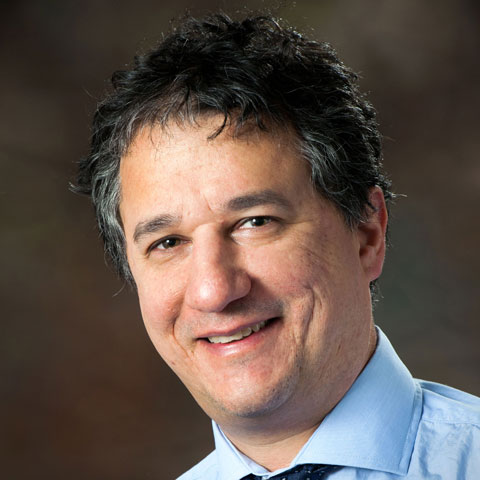
Dr. Nadine Fiani
Dr. Nadine Fiani is section chief and associate clinical professor in the Section of Dentistry and Oral Surgery at the Cornell University Hospital for Animals. She completed her BVSc at the University of Sydney and a residency in dentistry and oral surgery at the University of California at Davis. After some time in private practice, Fiani came to Cornell, where her clinical interests include endodontics, oncological surgery and maxillofacial trauma and surgical repair. Fiani researches clinical teaching measures of success, as well as the dentition of Australian mammals, etiological mechanisms of dental disease in zoo animals and endodontic disease. She is a diplomate of the American Veterinary Dental College.
Dr. Santiago Peralta
Dr. Santiago Peralta is an associate professor in the Section of Dentistry and Oral Surgery at the Cornell University Hospital for Animals. He received his degree in veterinary medicine from La Salle University in Bogota, Colombia, and worked in private practice for six years before specializing in dentistry and oral surgery. He completed a residency in veterinary dentistry and oral surgery at the University of California at Davis. Peralta then joined the faculty at Cornell, where his teaching emphasizes diagnostic and therapeutic principles of dental, oral and maxillofacial diseases of small animals. He is a diplomate of the American Veterinary Dental College and a Founding Fellow of Oral and Maxillofacial Surgery.
About the Cornell University College of Veterinary Medicine
The Cornell University College of Veterinary Medicine is dedicated to the creation, distribution and implementation of scientific knowledge to improve the health and well-being of animals and people. As a New York land-grant college, we achieve regional and global impact through education, discovery and care. Learn more about our history here.