


Foot Health
Introduction
Clinical lameness is a major cause of financial loss to dairy farmers due to milk loss, delayed conception, and costs related to extra handling, treatment, and early culling. By one estimate in New York herds, average economic loss is $350 per case. Additionally, lameness is a welfare concern and may indicate that facilities do not meet the comfort levels required for the cows. In the NYSCHAP Welfare Certification Program, the percent of visibly lame cattle is used as one factor in the assessment of facilities.
Surveys attempting to estimate the average prevalence of lameness range from 12–30 percent. The 2007 National Animal Health Monitoring System survey found that 94.9 percent of all dairy operations reported having at least one lame cow during 2006.
Additionally, producers identified 14.0 percent of their cows as lame during 2006. Research conducted by Cornell University, using local clinic data, estimates the prevalence of lameness to be closer to 30 percent. The 2007 National Market Cow and Bull Beef Quality Audit found that 49 percent of all dairy cows in holding pens at slaughter plants had a locomotion score greater than 1. The survey found only 16 percent of beef cows with a locomotion score greater than 1.
Early detection of lameness combined with a routine foot-trimming program is critical to minimize the impact on the farm. Lameness detection is also crucial for transport when marketing. Decisions for culling lame cows should be made expediently to ensure that cows remain ambulatory through the marketing chain. Farm personnel should be trained in a method of scoring animals for locomotion.
When used regularly, this tool can enable farms to detect lameness early and intervene before the condition worsens.
Although cows can be lame for a wide variety of reasons, 90 percent of the time the cause can be found by examining the foot. The conditions affecting the foot can result from infectious and/or noninfectious causes. This brochure discusses key causes and treatment and prevention of the common foot disorders that result in lameness and highlights prevention practices that can improve foot health.
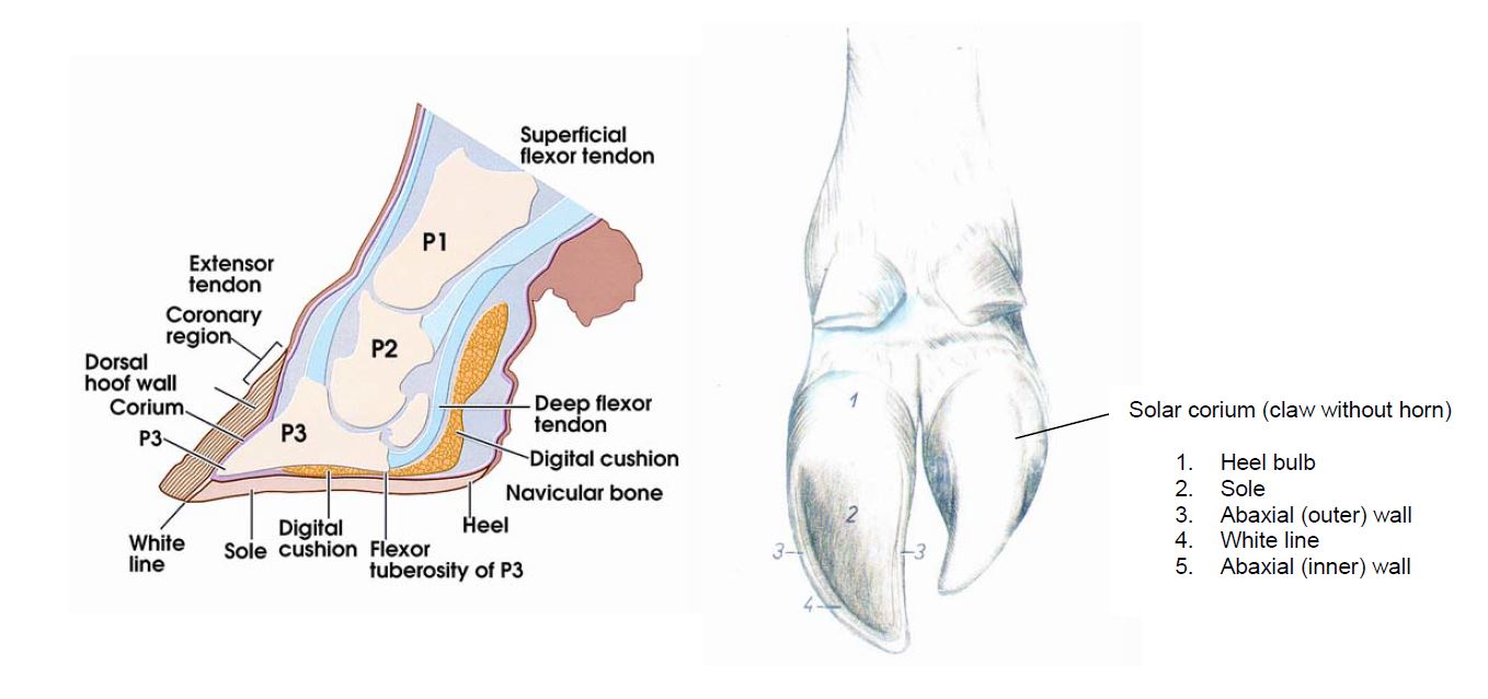
Foot Anatomy
Claw Trimming
Routine trimming is a critical component of lameness prevention. Corrective trimming removes excess horn growth and returns the claw to its normal shape, ensuring that weight is distributed evenly across the claw’s load-bearing structures. An overgrown toe places excessive pressure on the tendons and load is put onto the softer midsole region, which can lead to bruising, with a potential for ulceration.
Proper technique starts with knowledge of the normal shape for each claw. The so-called "Dutch Method" is a widely accepted method of claw trimming and follows six basic steps.* Restraint facilities should allow for efficient handling of cattle and prevent possible injury to cattle and farm personnel. Farm owners should consider owning their own foot-trimming table and tools to prevent entry of pathogens to the farm. Equipment must be well maintained and trimming tools must be sharp.
Lameness can result when the foot trimmer uses incorrect technique, trims too frequently, or trims just prior to moving cows to an area with abrasive flooring. Select a professional foot trimmer with proper training, experience, and demonstrated knowledge of foot care and lameness prevention. A foot trimmer should arrive with clean equipment, clothing, and tools. If a member of the farm plans to trim, make sure that person receives adequate training from a qualified professional. Blood-contaminated trimming equipment can spread blood-borne diseases such as bovine leukosis. In herds where this is a concern, blood-contaminated equipment should be disinfected between cows.
Optimal frequency of trimming varies by farm and depends on the rates of horn growth and wear. Most cows benefit from at least two trims per lactation—but cows should be individually assessed for the need for trimming. It is a good practice to inspect the feet of bred heifers and to trim if needed; those housed on bedded packs and in freestall barns may be especially prone to overgrown claws.
Laminitis
Description: Laminitis is an inflammation of the sensitive laminae and corium. Severely affected cattle will be acutely lame although many animals will be affected to a lesser degree. The effects of laminitis can be manifested in a variety of foot disorders that appear as: ridges along the foot wall; swelling at the coronary band; waxy, flaking solar horn tissue; false soles; hemorrhage in the sole; white line abscesses; and sole ulcers. All four feet are usually affected to some degree.
Cause (pathogenesis): Laminitis is caused by both nutritional and environmental components. The nutritional component of laminitis centers on a drop in pH of the rumen below approximately 5.8. This sets off a complex cascade of events that ultimately leads to a disruption in the sensitive laminae and corium of the foot and in many cases instability of the third phalanx. This drop in pH can be due to many factors, including insufficient amounts of rumination-enhancing fiber, excessive amounts of rumen-fermentable grain, slug-feeding patterns, sorting of the ration, major diet changes, and stress. The environmental components related to the incidence of laminitis center around excessive standing on concrete which can be related to uncomfortable stalls, over-crowding, heat stress, and long parlor-hold times (more than 3 hours/day).
Treatment: The underlying cause of laminitis occurrence should be addressed first. This should include a review of both the nutritional and environmental factors (as outlined above and below). Specific treatment at the foot level is aimed at correct trimming to reshape the claw so that weight is distributed appropriately across all weight-bearing surfaces. If there are specific lesions, such as white line abscesses or sole ulcers, these should be addressed as outlined in other sections of this pamphlet.
Prevention:
- Ensure that diets for lactating cows contain a minimum of 19 percent ADF and 28 percent NDF. Eight to 15 percent of the diet should be retained on the coarse screen of the Penn State particle separator and more than 50 percent retained on the top two screens.
- Assess feeding patterns to determine if there is evidence of sorting or slug-feeding.
- Optimize transition cow health and comfort, particularly in first-lactation heifers.
- Provide comfortable stalls that are appropriate to the size of the animals.
- Reduce overcrowding.
- Minimize the time cows spend in the parlor holding area and locked up for management purposes.
- Minimize stress and standing time
- Resurface floors that are slippery or potentially traumatic. Concrete floors should be grooved according to recommendations.*
- Rubber flooring can also be considered in heavily used areas.
- Implement routine foot trimming to maintain proper claw shape and redistribute weight appropriately across all weight-bearing surfaces.
- If possible, group first-lactation heifers together to minimize social stress.
- Minimize heat stress and fly exposure.
White Line Disease
Adapted from American Association of Bovine Practitioners (AABP) Lameness fact sheet "White Line Disease" (http://www.aabp.org)
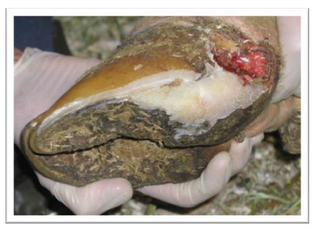
Description: White line disease (WLD) refers to a group of lesions that affect the junction between the sole and the wall of the claw. These lesions of the white line include fissures, hemorrhage (pinkness in the sole), and abscesses. The outer claw of the rear foot is most commonly affected.
Cause (pathogenesis): The lesions of WLD generally result from instability of the third digit within the claw horn capsule. This instability is thought to be the result of a complex cascade of events that may be triggered by hormonal changes surrounding calving, nutritional events such as subacute ruminal acidosis (SARA), or other changes in the rumen environment. When the animal bears weight, the loosened third digit causes further changes that lead to a weakening of the white line junction, especially in the location two-thirds of the way back from the toe on the outer claw. This can lead to separation of the foot wall from the sole, which then allows impaction of stones and other material. If this impaction is not relieved, it can lead to sepsis and abscess formation and may spread along the lamellae either to the coronary band or deeper into the foot.
Treatment: The objective is to functionally trim the claw, remove weight from the injured (usually outer) claw, and remove sufficient wall surrounding a lesion to expose healthy horn at the borders of the lesion. The heel of the affected claw should be trimmed level or lower than the sound claw, providing the sole does not become too thin. Affected claws should be blocked if this is not possible.
Severely affected animals should be housed on a bedded pack until they have recovered. Animals receiving a block should be reexamined in 4–5 weeks, with the block removed if healing has occurred, or replaced for an additional 4–5 weeks if more time is needed.
Prevention:
- Ensure that diets for lactating cows contain a minimum of 19 percent ADF and 28 percent NDF. Eight to 15 percent of the diet should be retained on the coarse screen of the Penn State particle separator and more than 50 percent retained on the top two screens.
- Assess feeding patterns to determine if there is evidence of sorting or slug-feeding.
- Optimize transition cow health and comfort, particularly in first-lactation heifers.
- Provide comfortable stalls that are appropriate to the size of the animals.
- Reduce overcrowding.
- Minimize the time cows spend in the parlor holding area and locked up for management purposes.
- Minimize stress and standing time
- Resurface floors that are slippery or potentially traumatic. Concrete floors should be grooved according to recommendations.* Rubber flooring can also be considered in heavily used areas.
- Implement routine foot trimming to maintain proper claw shape and redistribute weight appropriately across all weight-bearing surfaces.
- If possible, group first-lactation heifers together to minimize social stress.
- Minimize heat stress and fly exposure.
Sole Ulceration
Adapted from (AABP) Lameness fact sheet "Sole Ulceration" (http://www.aabp.org)
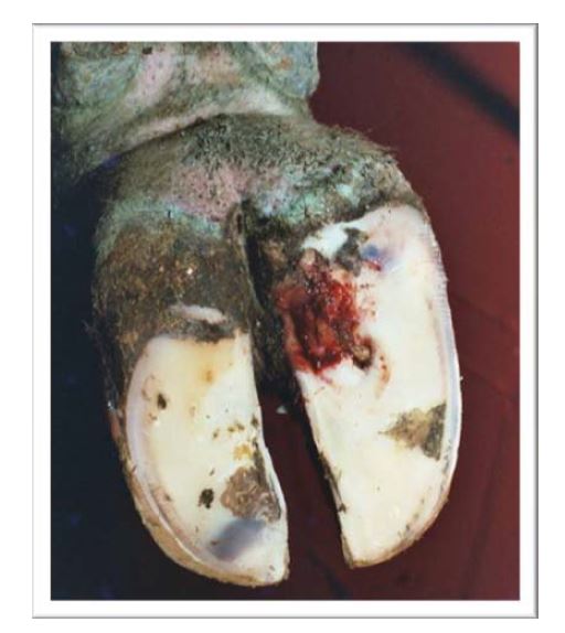
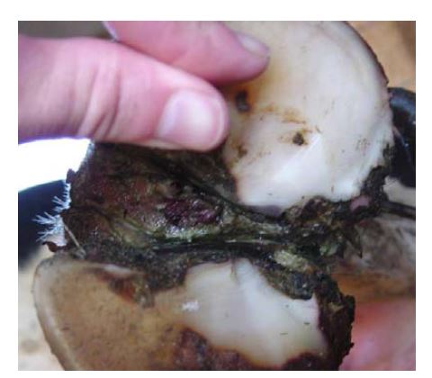
Description: A sole ulceration is a very painful, debilitating lesion that can lead to severe lameness. These ulcers are usually located in a very specific area on the sole (see picture below), although they can also be located in the heel or the toe. They are recognized as a focal area where very little to no horn remains to allow visualization of the solar corium. The solar corium in this situation is usually red in appearance, very sensitive, and may have formed granulation tissue causing it to protrude beyond the surrounding horn.
Cause (pathogenesis): Sole ulcers are caused by a disruption in normal horn producing
cells in a discrete area below the digital cushion due to pressure from an unstable third phalanx. This disruption in horn production eventually leads to a defect in the sole and protrusion of the solar corium. Instability of the third phalanx may result from hormonal changes at calving, nutritional events such as subacute ruminal acidosis (SARA), and severe cases of mastitis or metritis. Factors that promote overgrowth of the heel and sole, such as confinement on concrete walkways and standing surfaces, udder development, and abnormal confirmation, may predispose cows to ulcer formation. Improper trimming can also lead to abnormal weight-bearing and predispose to ulcer formation.
Treatment: The first step in treatment is to fully expose the ulcerated area by removing all loose horn tissue and thinning the margins of the ulcer. The second step is to transfer weight from the ulcer by functional claw trimming or applying a block on the sound claw. Any animal that has been blocked should be reexamined after approximately 4–5 weeks for retrimming, block removal, and assessment. If the ulcer is not fully healed at this point, a block should be reapplied. Bandaging the affected claw is not recommended in most cases, but animals should be segregated to a pasture or well-bedded area. Pain medication should be considered in these animals.
Prevention:
- Ensure that diets for lactating cows contain a minimum of 19 percent ADF and 28 percent NDF. Eight to 15 percent of the diet should be retained on the coarse screen of the Penn State particle separator and more than 50 percent retained on the top two screens.
- Assess feeding patterns to determine if there is evidence of sorting or slug-feeding.
- Optimize transition cow health and comfort, particularly in first-lactation heifers.
- Provide comfortable stalls that are appropriate to the size of the animals.
- Reduce overcrowding.
- Minimize the time cows spend in the parlor holding area and locked up for management purposes.
- Minimize stress and standing time.
- Resurface floors that are slippery or potentially traumatic. Concrete floors should be grooved according to recommendations.* Rubber flooring can also be considered in heavily used areas.
- Implement routine foot trimming to maintain proper claw shape and redistribute weight appropriately across all weight-bearing surfaces.
- If possible, group first-lactation heifers together to minimize social stress.
- Minimize heat stress and fly exposure.
-
Consider the following areas for herds that are experiencing a predominance of toe ulcers as compared to sole ulcers:
- reasons for rapid claw horn wear rates, such as abrasive surfaces
- trimming frequency
- trimming technique
Digital Dermatitis (Heel Warts)
Adapted from American Association of Bovine Practitioners (AABP) Lameness fact sheet "Digital Dermatitis (Heel Wart)" (http://www.aabp.org)
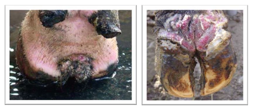
Description: Digital dermatitis (heel warts, strawberry heel warts, hairy heel warts) is an infectious disease that most commonly causes lesions on the rear foot in the heel region beginning at the cleft. Most lesions are between 1 and 2.5 inches in diameter and are circular or oval. More advanced lesions may form papillae that give the wart a "hairy" appearance. Lesions are painful and vary in color (red, black, gray, white-yellow, or combinations of colors). A foul odor may or may not be present.
Cause (pathogenesis): Digital dermatitis is a multifactorial disease that is most likely caused by infection from spirochetes of the genus Treponema. But it is clear that environment and management are large risk factors as well. Heel warts are commonly introduced to naive herds through purchased animals. Once introduced, the infection spreads rapidly throughout the herd. As the infection is established in the herd, older cows seem to develop immunity, as new infections are generally recognized in younger cows.
Infections are rarely seen in pastured cattle. New infections are commonly identified in fresh cows, suggesting that overall immune status is important. Herd infection is more common in free-stall environments than in tie-stall facilities. Environment of the farm is critically important in controlling the spread of disease. Decreasing the amount of time that cows stand in mud and manure helps to limit the amount of exposure to the organisms that cause heel warts. This can be influenced by cleanliness of the environment, as well as comfort of the stalls.
Treatment: Topical antibiotics are the most common treatment for heel warts. However, all are extra-label use and treatment records must be maintained. Foot sprays containing oxytetracycline or linomycin are effective and should be used according to veterinary direction. As well, a solution of either antibiotic may be soaked onto gauze and applied to the lesion. Other commercial nonantibiotic treatments may also be effective: consult with your veterinarian prior to using these. Foot baths are commonly used to prevent spread of infection within the herd.
Prevention:
- Keep all cow areas as clean and dry as possible and maintain cow comfort.
- Be certain that foot trimmers arrive with clean equipment and disinfect equipment between animals.
- Purchased cattle and cattle returning from custom heifer raisers or fairs can introduce heel warts. Quarantine cattle when possible and be aggressive in treating.
Interdigital Dermatitis
Anderson, D. E. (ed.) 2001. Diseases of the Digital Soft Tissues. in The Veterinary Clinics of North America: Food Animal Practice, xii+229 pp.
Description: This is a very common chronic infection of the skin around the claws and can be seen on many cows in New York State as "pitting" of the heels. In some cases this may be accompanied by heel cracks or heel horn erosion, and long term, the development of a fibroma (corn). Cows affected by this condition are not necessarily clinically lame. Unlike in foot rot, often all four feet are involved, although one foot may be favored over the rest. Older cows tend to show more evidence of this condition than younger ones.
Cause (pathogenesis): The primary causal agent is Dichelobacter nodosus, which thrives in humid, warm climates. These bacteria are carried by the cow on the skin, but the infection spreads from cow to cow through the environment, particularly through manure. It will not survive more than four days on the ground, but can persist in filth caked to the claws.
Treatment: For an animal that is clinically lame with interdigital dermatitis, trim away the excess tissue and reshape the foot closer to the correct shape.
Prevention: As with foot rot, constant wetness softens the skin and predisposes it to infection.
- Maintain clean alleyways and holding pens where cows are standing for prolonged periods of time.
- Trim feet regularly: this is a key part of preventing and controlling this disease.
- Use a well-maintained footbath to reduce the number of bacteria present on the foot and harden skin around the foot, making it more resistant to infection.
Use and Maintenance of Foot Baths
Foot baths and topical sprays are used to prevent spread of infectious diseases of the foot. Used correctly, sprays use less product and always deliver clean ingredient to the areas of the foot to which they are applied. However, sprays may not reach the areas of the foot that require coverage by the solution being used and in many cases a properly designed and managed foot bath will be a better option.
Producers should discuss with their herd veterinarian the type and concentration of solution to be used in the foot bath. Several active ingredients include copper sulfate, zinc sulfate, or formalin.* Antibiotics in footbaths are used in an extra-label fashion and should only be used upon prescription of the herd veterinarian. The frequency of use of foot baths will depend on the prevalence of infectious foot disease in the herd and, again, is a decision to be determined in consultation with the veterinarian.
Foot baths should be placed in an area where all animals needing treatment must walk through the bath in single file. The bath should be at least eight feet long and no more than 24 to 30 inches wide. Position the bath in an area where cow movement will not be impeded and where there will be no sharp corners for cows to navigate. The surface under the foot bath should be level and at the same height as the floor on which the cow is approaching the foot bath, so that animals do not have to step up or down when they walk through the foot bath. A pre-bath prior to the foot bath, which is used to remove manure from the feet of the animals, is recommended. The water-filled pre-bath should be the same dimensions as the foot bath and should be separated by at least six feet from the foot bath.
The level of solution in the pre-bath and foot bath should range from four to six inches and should be checked frequently for cleanliness of the solution. Dirty footbaths have the potential to worsen a herd problem. As a general rule, solutions should be changed in the baths after 150–200 cows have walked through them. However, this will depend on several factors, including whether a pre-bath is used and the severity of infectious foot disease in the herd.
Foot Rot (Interdigital Phlegmon)
Adapted from AABP fact sheet "Interdigital Phlegmon (Foot Rot)" and VCNA Food Animal Practice "Diseases of the Digital Soft Tissues"
Description: Foot rot is a bacterial infection of the interdigital space of the feet of dairy and beef cattle that results in rapid onset of severe lameness. There typically is symmetrical swelling just above both claws, necrosis of the skin in the interdigital space, and a foul smell associated with the lesion. Foot rot is commonly found in only one foot and is more common on rear feet.
Cause (pathogenesis): Foot rot is caused by specific bacteria that gain entry to the tissue of the foot via a skin defect. Trauma or conditions that predispose cattle to interdigital skin damage such as stones, stubble, pieces of wood, uneven ground, constant moisture, or dried mud are potential risk factors.
Treatment: Early detection is critical for successful treatment of foot rot. The interdigital space should be cleaned to identify this condition but bandaging is unnecessary. Systemic antibiotics are always indicated for a period of 3–5 days. Your veterinarian should be consulted as to which antibiotic is appropriate for your situation. There have been reports of "super foot-rot," which has resistance to some of the common antibiotic choices.
Prevention: Hygiene is the most important control measure. Reducing exposure to manure and avoiding chronic wetting of the foot are paramount to reducing the risk of foot rot.
- Frequently remove manure and maximize drainage from all areas of the facility.
- Maintain watering and feeding areas to avoid standing water and/or mud.
- Consider limiting access to known problem areas in pasture operations of beef and dairy cattle, or, if possible, only use historically problematic pastures during dry weather.
- Use a footbath with a range of disinfectants to clean and disinfect the interdigital skin.
- Provide dry places for cows to lie down.
*Gooch, C.A. 2003. Flooring Considerations for Dairy Cows. Natural Resource, Agriculture, and Engineering Service (NRAES) 148.