


In situ hybridization
In situ hybridization (ISH) is a technique that uses colorimetric or fluorescent probes to target and visualize specific DNA or RNA sequences within tissues. This technology can be broadly applied to study infectious agents, cancer, or developmental biology. Advanced Cell Diagnostics' RNAscope® ISH uses a proprietary probe design to target specific sequences of nucleic acid resulting in high specificity and a signal amplification system with enhanced sensitivity. In some cases, ISH can be more sensitive than PCR assay because it can label fragmented DNA or RNA. Moreover, ISH has the advantage of correlating the presence of infectious agents or tumor markers within lesions, and thus, provides a more definitive etiologic diagnosis.
ISH at the AHDC
We offer automated ISH services using RNAscope® probes designed and produced by Advanced Cell Diagnostics (ACD) for diagnostic applications using our state-of-the-art Ventana Discovery Ultra stainer. This allows consistent and rapid diagnostic or research output of reliable data. All ISH testing is performed using corresponding internal positive and negative controls to provide confidence in our findings. Interpretation of the results is provided by our board-certified anatomic pathologists with extensive RNAscope® ISH experience.
Currently, we offer a variety of probes targeting viruses or oncogenes associated with specific tumors. If your diagnostic or research needs require a probe that is not currently available on our list, an Anatomic Pathology Section representative will be happy to assist with your existing RNAscope® probe or developing a new probe with ACD.
Below is a list of probes currently available:
Bacteria
- Eubacteria
- Escherichia coli
Viruses
- Bovine papillomavirus types 1 and 2
- Canine herpesvirus
- Canine vesivirus
- Equine herpesvirus type 2
- Equine herpesvirus type 5
- Equine parvovirus-hepatitis
- West Nile virus
Tumor Diagnosis
- ETV1 marker for canine gastrointestinal stromal tumor
- Programmed death-ligand (PD-L1)
Submission Instructions for ISH
For each In-Situ Hybridization request, please provide either (I) a block of formalin-fixed and paraffin-embedded tissue (FFPE) or (II) four sections of FFPE tissue placed on positively charged, unstained glass slides prepared using a fresh microtome blade. Prepared slides should be shipped as quickly as possible and with ice packs to preserve the integrity of the nucleic acids in the sample. Samples submitted to the Animal Health Diagnostic Center must be accompanied by a completed submission form.
In situ hybridization provides a powerful tool for visualizing nucleic acids in tissues. Prolonged formalin fixation (more than a week), tissue autolysis, freeze-thawing, and other factors can adversely affect the integrity of nucleic acids. In our experience, ISH for DNA viruses is robust and can be used successfully in archived FFPE tissues that have been stored for over 20 years. If you have concerns about the quality of your sample, assessment of RNA integrity using ISH of species-specific housekeeping genes can be performed for an additional fee.
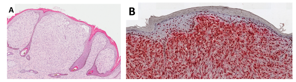
Detection of bovine papillomavirus type 1 and type 2 (BPV1/2) nucleic acid sequences by in-situ hybridization (ISH) of formalin-fixed and paraffin-embedded (FFPE) tissue section
Equine sarcoid and related soft tissue sarcomas
Differentiating equine sarcoids from soft tissue sarcomas (STSs) based on histomorphological features in routinely processed FFPE hematoxylin and eosin (H&E)-stained tissue sections can be challenging. Moreover, bovine papillomavirus type 1 and type 2 (BPV1/2) nucleic acid sequences, previously associated with sarcoids (Gaynor et al., 2016), have been found in certain dermal spindle cell tumors including 59% of peripheral nerve sheath tumors (PNSTs), 37% of fibrosarcomas, and 22% of other dermal spindle cell tumors of horses collectively referred as STSs (Epperson and Castleman, 2017). Therefore, BPV1/2 are associated with many STSs of horses in addition to sarcoids.
A highly sensitive and specific ISH assay has been validated for direct visualization of BPP1/2 nucleic acid sequences in routinely processed FFPE tissue sections and diagnosis of sarcoid and STSs in horses.
References
- Gaynor AM, Zhu KW, Dela Cruz FN Jr, et al. 2016. Localization of bovine papillomavirus nucleic acid in equine sarcoids. Veterinary Pathology 53:567-573.
- Epperson ED and Castleman WL. 2017. Bovine papillomavirus DNA and S100 profiles in sarcoids and other cutaneous spindle cell tumors in horses. Veterinary Pathology 54(1):44-52.
Bovine Fibropapilloma
A highly sensitive and specific ISH assay has been validated for direct visualization of BPP1/2 nucleic acids in routinely processed FFPE tissue sections and diagnostic confirmation of cutaneous and anogenital fibropapillomas in cattle.
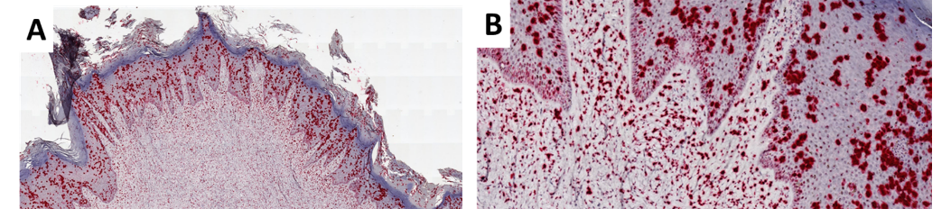
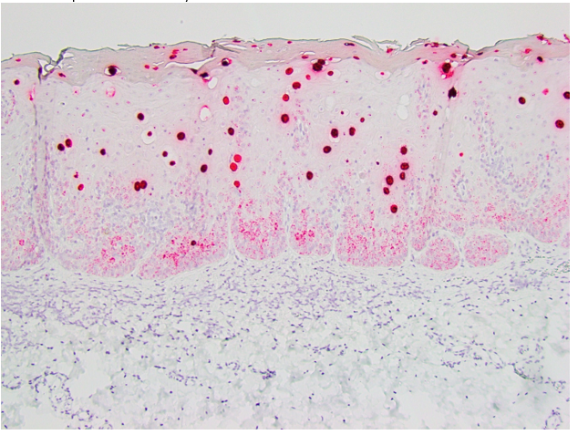
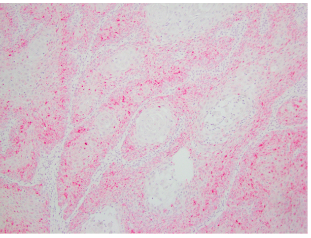
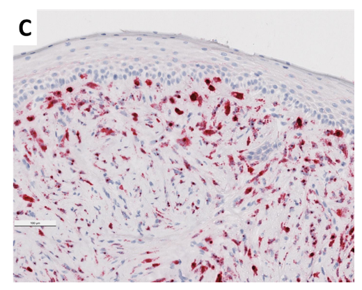 Photomicrographs of bovine cutaneous (A low and B high magnification) and genital (C) fibropapillomas ISH with Bovine Papillomavirus (BPV) type 1 and type 2 probe. Note prominent nuclear BPV1/2 signal within epidermal keratinocytes (A and B) and dermal fibroblast-like cells (A, B, and C) of bovine cutaneous (A and B) and genital (C) fibropapillomas.
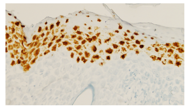
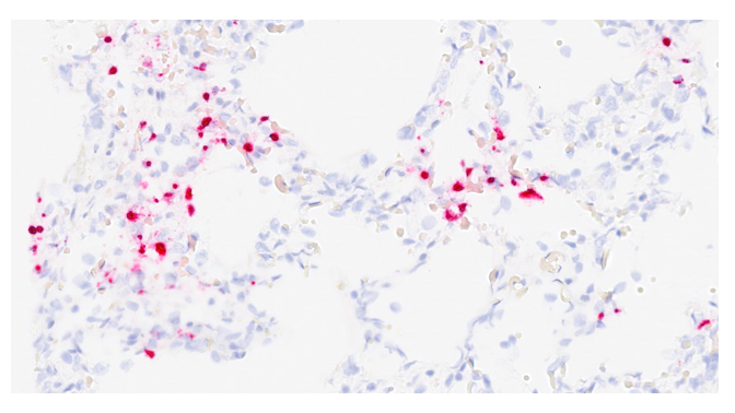
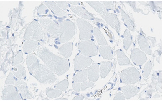
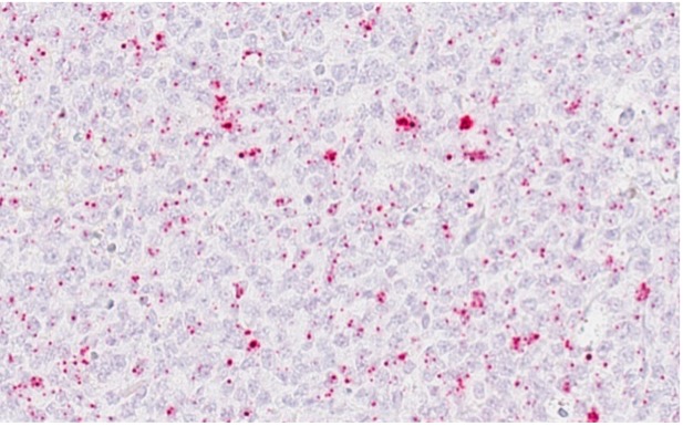
Detection of equine herpesvirus (EHV) type 5 nucleic acid sequence by in-situ hybridization (ISH) of formalin-fixed and paraffin-embedded (FFPE) tissue section
Equine Multinodular Pulmonary Fibrosis
An association between equine multinodular pulmonary fibrosis (EMPF) and the presence of equine gammaherpesvirus-5 (EHV-5) is well-established (Williams et al., 2007 and 2013; Marenzoni et al., 2011; Ochi et al., 2019). Horses with EMPF have EHV-5 nucleic acid primarily in pulmonary alveolar macrophages within fibrotic nodules.
A highly sensitive and specific ISH assay has been validated for direct visualization of EHV-5 nucleic acid sequences in routinely processed FFPE tissue sections and diagnostic confirmation of EMPF in horses.
References
- Marenzoni ML, Passamonti F, Lepr E, et al. 2011. Quantification of equid herpesvirus 5 DNA in clinical and necropsy specimens collected from a horse with equine multinodular pulmonary fibrosis. Journal of Veterinary Diagnostic Investigation 23:802-806.
- Williams KJ, Maes F, Del Piero F, et al. 2007. Equine multinodular pulmonary fibrosis: a newly recognized herpesvirus-associated fibrotic lung disease. Veterinary Pathology 44:849-862.
- Williams KJ, Robinson NE, Lim A, Brandenberg C, Maes R, Behan A, Bolin S. 2013. Experimental induction of pulmonary fibrosis in horses with the gammaherpesvirus equine herpesvirus 5. PLoS ONE 8(10): e77754.
- Ochi A, Sekiguchi M, Tsujimura K, et al., 2019. Two Cases of Equine Multinodular Pulmonary Fibrosis in Japan. Journal of Comparative Pathology 170:46-52.
Lymphohistiocytic Interface Dermatitis in Horses
Equid gammaherpesviruses 5 (EHV-5) has been associated with a non-painful, non-pruritic, scaly, annular to irregular muzzle lesion with histopathological features suggestive of either discoid lupus erythematosus-like disease (Cossic et al., 2017) or erythema multiforme (Herder et al., 2012). These lesions also display keratinocytes containing intranuclear inclusion bodies consistent with herpesvirus infection. A highly sensitive and specific ISH assay has been validated for direct visualization of EHV-5 nucleic acid sequences in routinely processed FFPE skin sections and diagnostic confirmation of EHV-5 associated dermatitis in horses.
References
- Herder V, Barsnick R, Walliser U, et al., 2012. Equid herpesvirus 5-associated dermatitis in a horse--resembling herpes-associated erythema multiforme. Veterinary Microbiology 155(2-4):420-424.
- Cossic B, Glaser AL, Duhamel GE, et al., 2017. Association of equine gammaherpesvirus-5 with lymphocytic interface dermatitis on the muzzle of two horses in the United States. American College of Veterinary Pathologists Annual Meeting, Vancouver, Canada, November 4-8.
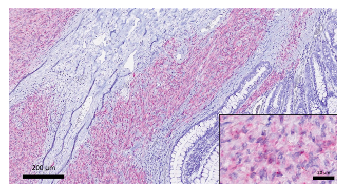
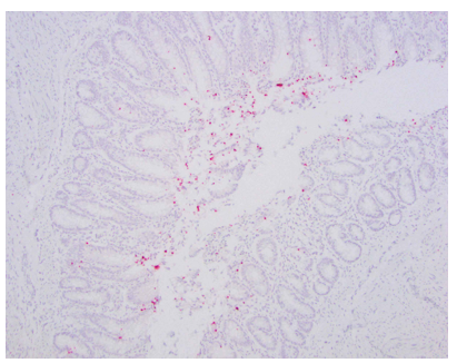
Detection of equine herpesvirus (EHV) type 2 and 5 nucleic acid sequences by in-situ hybridization (ISH) of formalin-fixed and paraffin-embedded (FFPE) gastric tissue from horses with Equine Gastric Ulcer Syndrome
Equine Gastric Ulcer Syndrome
Equid gammaherpesviruses 2 and 5 (EHV-2 and -5) are commonly seen within gastric mucosal epithelium of horses where they may contribute to equine gastric ulcer syndrome (EGUS).
Highly sensitive and specific ISH assays have been validated for direct visualization of EHV-2 and EHV-5 nucleic acid sequences in routinely processed FFPE tissue sections and diagnostic confirmation of EHV-associated EGUS in horses.
Reference
- Pennington MR, Cossic BGA, Perkins GA, et al., 2017. First demonstration of equid gammaherpesviruses within the gastric mucosal epithelium of horses. Virus Research 242:30-36.

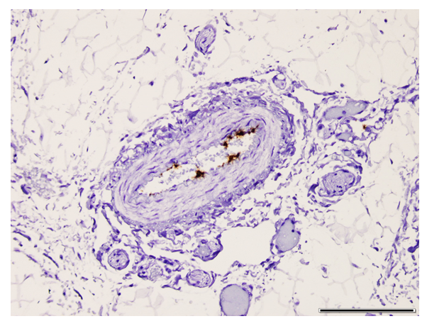
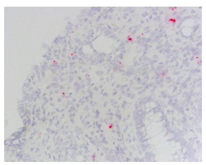
Detection of Canine Vesivirus (CaVV) nucleic acid sequence by in-situ hybridization (ISH) in formalin-fixed and paraffin-embedded (FFPE) tissue sections
Canine Vesivirus
A novel systemic fatal disease of dogs with hemorrhagic gastroenteritis has been associated with the presence of canine vesivirus (CaVV), a genus in the Caliciviridae family (Renshaw et al., 2018). Histologically, the disease is characterized by a generalized vasculopathy and CaVV nucleic acids signal within endothelial cells of arterial blood vessels and capillaries. CaVV nucleic acids signal is associated with necrosis and hemorrhage, primarily within the intestinal tract, but also in glial cells and neurons of the brain, circulating leukocytes, mononuclear cells in lymph nodes, spleen red pulp, intestinal lamina propria, and bone marrow of affected dogs.
A highly sensitive and specific ISH assay has been validated for direct visualization of CaVV nucleic acid sequences in routinely processed FFPE tissue sections and diagnostic confirmation of infection in dogs.
Reference
- Renshaw RW, Griffing J, Weisman J, Crofton LM, Laverack MA, Poston RP, Duhamel GE, Dubovi EJ. 2018. Characterization of a Vesivirus associated with an outbreak of acute hemorrhagic gastroenteritis in domestic dogs. J Clin Microbiol 56(5):e01951-17.
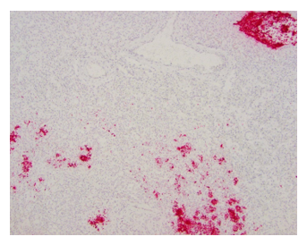
Detection of E26 Transformation-specific sequent variant 1 (ETV1) mRNA expression in Canine Cecal Gastrointestinal Stromal Tumors (GIST) by in-situ hybridization (ISH) in formalin-fixed and paraffin-embedded (FFPE) tissue sections
Canine Cecal Gastrointestinal Stromal Tumors (GIST)
Detection of ETV1 mRNA expression by ISH is a useful diagnostic marker of gastrointestinal stromal tumor (GIST) in humans, particularly when immunohistochemical expression of CD117 (c-kit) and DOG1 markers is lost (Jang et al., 2015). A highly sensitive and specific ISH assay has been validated for direct visualization of ETV1 nucleic acid sequences in routinely processed FFPE tissue sections and diagnostic confirmation of GIST in dogs.
References
- Jang BG, Lee HE, Kim WH. 2015. ETV1 mRNA is specifically expressed in gastrointestinal stromal tumors. Virchow Arch 467(4):393-403.
- Cavasin JP and Duhamel GE. 2020. Detection of E26 transformation-specific sequence variant 1 (ETV1) mRNA expression in canine cecal gastrointestinal stromal tumors by in-situ hybridization. American College of Veterinary Pathologists Annual Meeting.
Detection of Canine Herpesvirus (CHV) nucleic acid sequence by in-situ hybridization (ISH) in formalin-fixed and paraffin-embedded (FFPE) tissue sections
Canine Herpesvirus
Detection of CHV mRNA expression by ISH is a useful diagnostic marker of infection. While this disease presents with characteristic gross lesions, CHV can be difficult to identify in adult infections and in neonatal cases in which gross lesions are not readily identifiable.
A highly sensitive and specific ISH assay has been validated for direct visualization of CHV nucleic acid sequences in routinely processed FFPE tissue sections and diagnostic confirmation of CHV infection in dogs.
Reference
- Jager M, Sloma EA, Shelton M, Miller AD. 2017. Characterization of naturally acquired canine herpesvirus-associated meningoencephalitis. Vet Pathol 54(5):820-827.
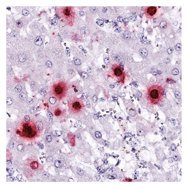
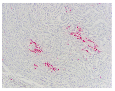
Detection of Equine Parvovirus-Hepatitis (EqPV-H) nucleic acid sequence by in-situ hybridization (ISH) in formalin-fixed and paraffin-embedded (FFPE) tissue sections
Equine Parvovirus-Hepatitis
Equine parvovirus-hepatitis (EqPV-H) causes hepatitis ranging in severity from subclinical liver enzyme elevations to fulminant liver necrosis (Theiler’s disease)1–6. The virus is transmitted naturally between horses on the same farm or iatrogenically through the administration of equine biologics such as tetanus antitoxin and allogenic stem cell therapy1,3. The primary mode of transmission is currently unknown. Currently, testing options for EqPV-H include PCR and ISH. A highly sensitive and specific ISH assay has been validated for direct visualization of EqPV-H nucleic acid sequences in routinely processed FFPE tissue sections and diagnostic confirmation of EqPV-H infection in horses. This useful tool can confirm the presence of the pathogen within affected tissues. Horses can persistently test EqPV-H PCR positive long after hepatitis has resolved, resulting in difficult interpretation of positive PCR results. ISH should be considered when a horse has serum chemistry changes consistent with hepatitis and tests positive for EqPV-H on PCR of serum or liver, when a liver biopsy reveals hepatitis, and in cases of fulminant hepatic necrosis.
References
- Tomlinson, J. E. et al. Tropism, pathology, and transmission of equine parvovirus-hepatitis. Emerging Microbes & Infections 9, 651–663 (2020).
- Vengust, M. et al. First report of equine parvovirus-hepatitis-associated Theiler’s disease in Europe. Equine Veterinary Journal n/a,.
- Tomlinson, J. E. et al. Viral testing of 18 consecutive cases of equine serum hepatitis: A prospective study (2014-2018). Journal of Veterinary Internal Medicine 33, 251–257 (2019).
- Tomlinson, J. E. et al. Viral testing of 10 cases of Theiler’s disease and 37 in-contact horses in the absence of equine biologic product administration: A prospective study (2014-2018). Journal of Veterinary Internal Medicine 33, 258–265 (2019).
- Tomlinson, J. E., Van de Walle, G. R. & Divers, T. J. What Do We Know About Hepatitis Viruses in Horses? Veterinary Clinics of North America: Equine Practice S0749073919300136 (2019) doi:10.1016/j.cveq.2019.03.001.
- Divers, T. J. et al. New Parvovirus Associated with Serum Hepatitis in Horses after Inoculation of Common Biological Product. Emerging Infectious Diseases 24, 303–310 (2018).
Detection of Escherichia coli nucleic acid sequence by in-situ hybridization (ISH) in formalin-fixed and paraffin-embedded (FFPE) tissue sections
Escherichia coli mRNA
Detection of Escherichia coli mRNA expression by ISH is a useful diagnostic marker of infection and can be used in cases in which culture was not obtained or is unavailable. It can also be used in cases of histiocytic ulcerative colitis (of Boxer and Bulldogs) to determine the presence of the bacterium.
A highly sensitive and specific ISH assay has been validated for diagnostic confirmation of infection through direct visualization of E. coli nucleic acid sequences in routinely processed FFPE tissue sections.
Detection of eubacterial nucleic acid sequence by in-situ hybridization (ISH) in formalin-fixed and paraffin-embedded (FFPE) tissue sections
Eubacterial Nucleic Acid
Detection of eubacterial mRNA expression by ISH is a useful diagnostic marker of the presence of eubacterial cells and can be used to supplement cases in which culture is not available or determining the presence of invasive bacteria (i.e. colonic invasion of bacteria.)
A highly sensitive and specific ISH assay has been validated for direct visualization of eubacterial nucleic acid sequences in routinely processed FFPE tissue sections and diagnostic confirmation of infection.
Detection of West Nile virus (WNV) nucleic acid sequence by in-situ hybridization (ISH) in formalin-fixed and paraffin-embedded (FFPE) tissue sections
West Nile virus (WNV)
Detection of West Nile virus (WNV) mRNA expression by ISH is a useful diagnostic marker of the presence of WNV infection and offers a more sensitive assay than immunohistochemistry.
A highly sensitive and specific ISH assay has been validated for direct visualization of WNV nucleic acid sequences in routinely processed FFPE tissue sections and diagnostic confirmation of infection.
Detection of Equus caballus papillomavirus 2 (EcPV2) nucleic acid sequence by in-situ hybridization (ISH) in formalin-fixed and paraffin-embedded (FFPE) tissue sections
Equus caballus papillomavirus 2 (EcPV2)
A subset of equine genital and gastric squamous cell carcinomas are associated with Equus caballus papillomavirus-2 (EcPV2) infection. A highly sensitive and specific ISH assay has been validated for direct visualization of EcPV2 nucleic acid sequences in routinely processed FFPE tissue sections and diagnostic confirmation of infection.
Detection of Programmed death-ligand (PD-L1) nucleic acid sequence by in-situ hybridization (ISH) in formalin-fixed and paraffin-embedded (FFPE) tissue sections
Programmed death-ligand 1 (PD-L1)
Programmed death-ligand 1 (PD-L1) binds to the inhibitory checkpoint molecule PD-1. Upregulation of PD-L1 is thought to allow neoplastic cells to evade the host immune system. PD-L1 is highly expressed in a variety of canine malignancies, including squamous cell carcinoma, nasal adenocarcinoma, urothelial (transitional) cell carcinoma, carcinoma of the apocrine glands of the anal sac, osteosarcoma, oral malignant melanoma, mammary adenocarcinoma, histiocytic sarcoma and diffuse large B-cell lymphoma (1). Analysis of PD-L1 expression in human cancer is employed as a prognostic marker. For example, high expression on renal cell carcinoma is associated with increased tumor aggressiveness and a 4.5 fold increased risk of death (2). High expression is used to identify candidates for immunotherapy with PD-L1 inhibitory monoclonal antibodies (eg. duravalumab, atezolizumab and avelumab). Promising results with the use of inhibitory PD-L1 monoclonal antibody therapy have been reported in dogs (1).

References
- Maekawa N, et al. PD-L1 immunohistochemistry for canine cancers and clinical benefit of anti-PD-L1 antibody in dogs with pulmonary metastatic oral malignant melanoma. NPJ Precis Oncol 5:10, 2021. Doi: 10.1038/s41698-021-00147-6
- Thompson RH, Gillett MD, Cheville JC, Lohse CM, Dong H, Webster WS, et al"Costimulatory B7-H1 in renal cell carcinoma patients: Indicator of tumor aggressiveness and potential therapeutic target". PNAS USA 101 (49): 17174–17179, 2004.