


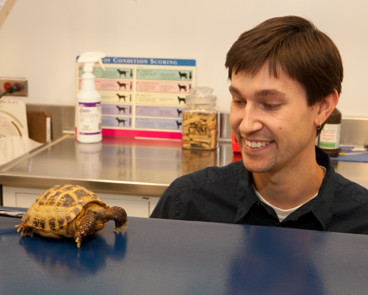
Companion Animal Hospital
Imaging Service

The Imaging Service at the Cornell University Hospital for Animals takes pride in providing excellent patient care and customer service.
Emergency and Critical Care
The Emergency and Critical Care Service at the Cornell University Hospital for Animals provides evaluation, medical care and surgical treatment to severely injured or ill companion animals, as well as ongoing care for critically ill or injured animals 24 hours a day, 365 days a year.
Anesthesia and Pain Medicine
Anesthesiology services
Cornell’s Anesthesiology and Pain Medicine service pioneers the development and clinical application of ultrasound guided regional anesthesia techniques, as well as cardiopulmonary monitoring. We collaborate closely with your pet's primary care team to develop individualized treatment plans. Additionally, we offer chronic pain management services, including interventional techniques like epidural, peripheral nerve, and joint injections.